6MD6

CRYSTAL STRUCTURE ANALYSIS OF PLANT EXOHYDROLASE IN COMPLEX WITH METHYL 2-THIO-BETA-SOPHOROSIDE


Experimental Data Snapshot

  • Method: X-RAY DIFFRACTION
  • Resolution: 1.68 Å
  • R-Value Free: 0.159 
  • R-Value Work: 0.132 
  • R-Value Observed: 0.133 

Starting Model: experimental
View more details

wwPDB Validation   3D Report Full Report


Ligand Structure Quality Assessment 


This is version 2.1 of the entry. See complete history


Literature

Discovery of processive catalysis by an exo-hydrolase with a pocket-shaped active site.

Streltsov, V.A.Luang, S.Peisley, A.Varghese, J.N.Ketudat Cairns, J.R.Fort, S.Hijnen, M.Tvaroska, I.Arda, A.Jimenez-Barbero, J.Alfonso-Prieto, M.Rovira, C.Mendoza, F.Tiessler-Sala, L.Sanchez-Aparicio, J.E.Rodriguez-Guerra, J.Lluch, J.M.Marechal, J.D.Masgrau, L.Hrmova, M.

(2019) Nat Commun 10: 2222-2222

  • DOI: https://doi.org/10.1038/s41467-019-09691-z
  • Primary Citation of Related Structures:  
    3WLH, 3WLI, 3WLJ, 3WLK, 3WLL, 3WLM, 3WLN, 3WLO, 3WLP, 3WLQ, 3WLR, 6MD6, 6MI1

  • PubMed Abstract: 

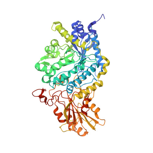
    Substrates associate and products dissociate from enzyme catalytic sites rapidly, which hampers investigations of their trajectories. The high-resolution structure of the native Hordeum exo-hydrolase HvExoI isolated from seedlings reveals that non-covalently trapped glucose forms a stable enzyme-product complex. Here, we report that the alkyl β-D-glucoside and methyl 6-thio-β-gentiobioside substrate analogues perfused in crystalline HvExoI bind across the catalytic site after they displace glucose, while methyl 2-thio-β-sophoroside attaches nearby. Structural analyses and multi-scale molecular modelling of nanoscale reactant movements in HvExoI reveal that upon productive binding of incoming substrates, the glucose product modifies its binding patterns and evokes the formation of a transient lateral cavity, which serves as a conduit for glucose departure to allow for the next catalytic round. This path enables substrate-product assisted processive catalysis through multiple hydrolytic events without HvExoI losing contact with oligo- or polymeric substrates. We anticipate that such enzyme plasticity could be prevalent among exo-hydrolases.


  • Organizational Affiliation

    Commonwealth Scientific and Industrial Research Organisation, Materials Science and Engineering, Parkville Victoria, 3052, Australia.


Macromolecules
Find similar proteins by:  (by identity cutoff)  |  3D Structure
Entity ID: 1
MoleculeChains Sequence LengthOrganismDetailsImage
Beta-D-glucan exohydrolase isoenzyme ExoI609Hordeum vulgare subsp. vulgareMutation(s): 1 
UniProt
Find proteins for Q9XEI3 (Hordeum vulgare subsp. vulgare)
Explore Q9XEI3 
Go to UniProtKB:  Q9XEI3
Entity Groups  
Sequence Clusters30% Identity50% Identity70% Identity90% Identity95% Identity100% Identity
UniProt GroupQ9XEI3
Glycosylation
Glycosylation Sites: 3
Sequence Annotations
Expand
  • Reference Sequence
Oligosaccharides

Help

Entity ID: 2
MoleculeChains Length2D Diagram Glycosylation3D Interactions
beta-D-glucopyranose-(1-2)-methyl 2-thio-beta-D-glucopyranoside
B
2N/AN/A
Glycosylation Resources
GlyTouCan:  G67616SG
GlyCosmos:  G67616SG
Small Molecules
Ligands 4 Unique
IDChains Name / Formula / InChI Key2D Diagram3D Interactions
1PE
Query on 1PE

Download Ideal Coordinates CCD File 
R [auth A]
S [auth A]
T [auth A]
U [auth A]
V [auth A]
R [auth A],
S [auth A],
T [auth A],
U [auth A],
V [auth A],
W [auth A]
PENTAETHYLENE GLYCOL
C10 H22 O6
JLFNLZLINWHATN-UHFFFAOYSA-N
NAG
Query on NAG

Download Ideal Coordinates CCD File 
C [auth A],
D [auth A],
E [auth A]
2-acetamido-2-deoxy-beta-D-glucopyranose
C8 H15 N O6
OVRNDRQMDRJTHS-FMDGEEDCSA-N
SO4
Query on SO4

Download Ideal Coordinates CCD File 
P [auth A],
Q [auth A]
SULFATE ION
O4 S
QAOWNCQODCNURD-UHFFFAOYSA-L
GOL
Query on GOL

Download Ideal Coordinates CCD File 
F [auth A]
G [auth A]
H [auth A]
I [auth A]
J [auth A]
F [auth A],
G [auth A],
H [auth A],
I [auth A],
J [auth A],
K [auth A],
L [auth A],
M [auth A],
N [auth A],
O [auth A]
GLYCEROL
C3 H8 O3
PEDCQBHIVMGVHV-UHFFFAOYSA-N
Biologically Interesting Molecules (External Reference) 1 Unique
Experimental Data & Validation

Experimental Data

  • Method: X-RAY DIFFRACTION
  • Resolution: 1.68 Å
  • R-Value Free: 0.159 
  • R-Value Work: 0.132 
  • R-Value Observed: 0.133 
  • Space Group: P 43 21 2
Unit Cell:
Length ( Å )Angle ( ˚ )
a = 100.311α = 90
b = 100.311β = 90
c = 182.153γ = 90
Software Package:
Software NamePurpose
REFMACrefinement
XDSdata reduction
Aimlessdata scaling
MOLREPphasing

Structure Validation

View Full Validation Report


Ligand Structure Quality Assessment 


Entry History 

Deposition Data

Revision History  (Full details and data files)

  • Version 1.0: 2019-05-29
    Type: Initial release
  • Version 1.1: 2019-06-05
    Changes: Data collection, Database references
  • Version 2.0: 2020-07-29
    Type: Remediation
    Reason: Carbohydrate remediation
    Changes: Atomic model, Data collection, Derived calculations, Non-polymer description, Structure summary
  • Version 2.1: 2023-10-11
    Changes: Data collection, Database references, Refinement description, Structure summary