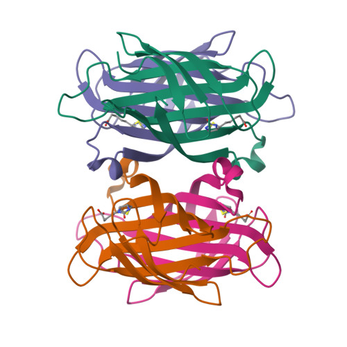
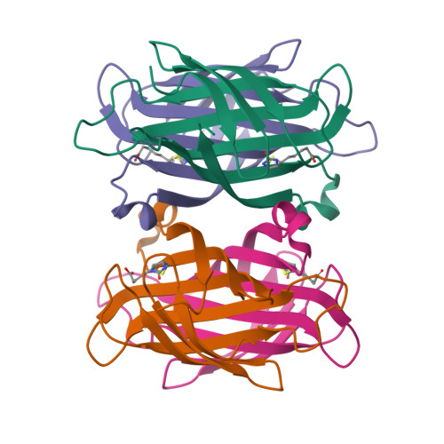
6M9B

Wild-type streptavidin in complex with biotin solved by native SAD with data collected at 6 keV


Experimental Data Snapshot

  • Method: X-RAY DIFFRACTION
  • Resolution: 1.55 Å
  • R-Value Free: 0.183 
  • R-Value Work: 0.154 
  • R-Value Observed: 0.155 

wwPDB Validation   3D Report Full Report


Ligand Structure Quality Assessment 


This is version 1.2 of the entry. See complete history


Literature

Making routine native SAD a reality: lessons from beamline X06DA at the Swiss Light Source.

Basu, S.Finke, A.Vera, L.Wang, M.Olieric, V.

(2019) Acta Crystallogr D Struct Biol 75: 262-271

  • DOI: https://doi.org/10.1107/S2059798319003103
  • Primary Citation of Related Structures:  
    6M9B

  • PubMed Abstract: 

    Native single-wavelength anomalous dispersion (SAD) is the most attractive de novo phasing method in macromolecular crystallography, as it directly utilizes intrinsic anomalous scattering from native crystals. However, the success of such an experiment depends on accurate measurements of the reflection intensities and therefore on careful data-collection protocols. Here, the low-dose, multiple-orientation data-collection protocol for native SAD phasing developed at beamline X06DA (PXIII) at the Swiss Light Source is reviewed, and its usage over the last four years on conventional crystals (>50 µm) is reported. Being experimentally very simple and fast, this method has gained popularity and has delivered 45 de novo structures to date (13 of which have been published). Native SAD is currently the primary choice for experimental phasing among X06DA users. The method can address challenging cases: here, native SAD phasing performed on a streptavidin-biotin crystal with P2 1 symmetry and a low Bijvoet ratio of 0.6% is highlighted. The use of intrinsic anomalous signals as sequence markers for model building and the assignment of ions is also briefly described.


  • Organizational Affiliation

    Swiss Light Source, Paul Scherrer Institut, Villigen PSI, Switzerland.


Macromolecules
Find similar proteins by:  (by identity cutoff)  |  3D Structure
Entity ID: 1
MoleculeChains Sequence LengthOrganismDetailsImage
Streptavidin
A, B, C, D
127Streptomyces avidiniiMutation(s): 0 
UniProt
Find proteins for P22629 (Streptomyces avidinii)
Explore P22629 
Go to UniProtKB:  P22629
Entity Groups  
Sequence Clusters30% Identity50% Identity70% Identity90% Identity95% Identity100% Identity
UniProt GroupP22629
Sequence Annotations
Expand
  • Reference Sequence
Experimental Data & Validation

Experimental Data

  • Method: X-RAY DIFFRACTION
  • Resolution: 1.55 Å
  • R-Value Free: 0.183 
  • R-Value Work: 0.154 
  • R-Value Observed: 0.155 
  • Space Group: P 1 21 1
Unit Cell:
Length ( Å )Angle ( ˚ )
a = 50.978α = 90
b = 98.322β = 112.64
c = 52.795γ = 90
Software Package:
Software NamePurpose
PHENIXrefinement
XDSdata reduction
XSCALEdata scaling
SHARPphasing

Structure Validation

View Full Validation Report


Ligand Structure Quality Assessment 


Entry History 

Deposition Data

  • Released Date: 2019-04-17 
  • Deposition Author(s): Finke, A.D.

Revision History  (Full details and data files)

  • Version 1.0: 2019-04-17
    Type: Initial release
  • Version 1.1: 2019-11-13
    Changes: Database references
  • Version 1.2: 2024-03-13
    Changes: Data collection, Database references