6M83 | pdb_00006m83

Crystal structure of TylM1 S120A bound to SAH and dTDP-phenol


Experimental Data Snapshot

  • Method: X-RAY DIFFRACTION
  • Resolution: 1.37 Å
  • R-Value Free: 
    0.182 (Depositor), 0.182 (DCC) 
  • R-Value Work: 
    0.153 (Depositor), 0.154 (DCC) 
  • R-Value Observed: 
    0.154 (Depositor) 

wwPDB Validation 3D Report Full Report

Validation slider image for 6M83

Ligand Structure Quality Assessment 


This is version 1.3 of the entry. See complete history

Literature

Structural and Functional Characterization of Sulfonium Carbon-Oxygen Hydrogen Bonding in the Deoxyamino Sugar Methyltransferase TylM1.

Fick, R.J.Horowitz, S.McDole, B.G.Clay, M.C.Mehl, R.A.Al-Hashimi, H.M.Scheiner, S.Trievel, R.C.

(2019) Biochemistry 58: 2152-2159

  • DOI: https://doi.org/10.1021/acs.biochem.8b01141
  • Primary Citation Related Structures: 
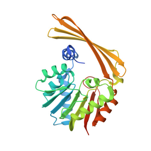
    6M81, 6M82, 6M83

  • PubMed Abstract: 

    The N-methyltransferase TylM1 from Streptomyces fradiae catalyzes the final step in the biosynthesis of the deoxyamino sugar mycaminose, a substituent of the antibiotic tylosin. The high-resolution crystal structure of TylM1 bound to the methyl donor S-adenosylmethionine (AdoMet) illustrates a network of carbon-oxygen (CH···O) hydrogen bonds between the substrate's sulfonium cation and residues within the active site. These interactions include hydrogen bonds between the methyl and methylene groups of the AdoMet sulfonium cation and the hydroxyl groups of Tyr14 and Ser120 in the enzyme. To examine the functions of these interactions, we generated Tyr14 to phenylalanine (Y14F) and Ser120 to alanine (S120A) mutations to selectively ablate the CH···O hydrogen bonding to AdoMet. The TylM1 S120A mutant exhibited a modest decrease in its catalytic efficiency relative to that of the wild type (WT) enzyme, whereas the Y14F mutation resulted in an approximately 30-fold decrease in catalytic efficiency. In contrast, site-specific substitution of Tyr14 by the noncanonical amino acid p-aminophenylalanine partially restored activity comparable to that of the WT enzyme. Correlatively, quantum mechanical calculations of the activation barrier energies of WT TylM1 and the Tyr14 mutants suggest that substitutions that abrogate hydrogen bonding with the AdoMet methyl group impair methyl transfer. Together, these results offer insights into roles of CH···O hydrogen bonding in modulating the catalytic efficiency of TylM1.


  • Organizational Affiliation
    • Department of Biological Chemistry , University of Michigan , Ann Arbor , Michigan 48109 , United States.

Macromolecule Content 

  • Total Structure Weight: 29.42 kDa 
  • Atom Count: 2,103 
  • Modeled Residue Count: 237 
  • Deposited Residue Count: 263 
  • Unique protein chains: 1

Macromolecules

Find similar proteins by:|  3D Structure
Entity ID: 1
MoleculeChains  Sequence LengthOrganismDetailsImage
dTDP-3-amino-3,6-dideoxy-alpha-D-glucopyranose N,N-dimethyltransferase263Streptomyces fradiaeMutation(s): 1 
Gene Names: tylM1tylMI
EC: 2.1.1.235
UniProt
Find proteins for P95748 (Streptomyces fradiae)
Explore P95748 
Go to UniProtKB:  P95748
Entity Groups
Sequence Clusters30% Identity50% Identity70% Identity90% Identity95% Identity100% Identity
UniProt GroupP95748
Sequence Annotations
Expand
Reference Sequence

Small Molecules

Ligands 3 Unique
IDChains Name / Formula / InChI Key2D Diagram3D Interactions
TLO

Query on TLO


Download:Ideal Coordinates CCD File
C [auth A]5'-O-[(S)-hydroxy{[(S)-hydroxy(phenoxy)phosphoryl]oxy}phosphoryl]thymidine
C16 H20 N2 O11 P2
BANJVYKYIQPYKP-BFHYXJOUSA-N
SAH

Query on SAH


Download:Ideal Coordinates CCD File
B [auth A]S-ADENOSYL-L-HOMOCYSTEINE
C14 H20 N6 O5 S
ZJUKTBDSGOFHSH-WFMPWKQPSA-N
CL

Query on CL


Download:Ideal Coordinates CCD File
D [auth A]CHLORIDE ION
Cl
VEXZGXHMUGYJMC-UHFFFAOYSA-M

Experimental Data & Validation

Experimental Data

  • Method: X-RAY DIFFRACTION
  • Resolution: 1.37 Å
  • R-Value Free:  0.182 (Depositor), 0.182 (DCC) 
  • R-Value Work:  0.153 (Depositor), 0.154 (DCC) 
  • R-Value Observed: 0.154 (Depositor) 
Space Group: I 2 2 2
Unit Cell:
Length ( Å )Angle ( ˚ )
a = 64.521α = 90
b = 91.267β = 90
c = 93.629γ = 90
Software Package:
Software NamePurpose
HKL-2000data reduction
HKL-2000data scaling
PHASERphasing
PHENIXrefinement
PDB_EXTRACTdata extraction

Structure Validation

View Full Validation Report


Ligand Structure Quality Assessment 


Entry History 

& Funding Information

Deposition Data


Funding OrganizationLocationGrant Number
National Science Foundation (NSF, United States)United StatesCHE-1508492

Revision History  (Full details and data files)

  • Version 1.0: 2019-03-13
    Type: Initial release
  • Version 1.1: 2019-05-01
    Changes: Data collection, Database references
  • Version 1.2: 2019-11-27
    Changes: Author supporting evidence
  • Version 1.3: 2024-03-13
    Changes: Data collection, Database references