6M63 | pdb_00006m63

Crystal structure of a cAMP sensor G-Flamp1.


Experimental Data Snapshot

  • Method: X-RAY DIFFRACTION
  • Resolution: 2.25 Å
  • R-Value Free: 
    0.223 (Depositor), 0.224 (DCC) 
  • R-Value Work: 
    0.178 (Depositor), 0.180 (DCC) 
  • R-Value Observed: 
    0.180 (Depositor) 

Starting Models: experimental
View more details

wwPDB Validation 3D Report Full Report

Validation slider image for 6M63

Ligand Structure Quality Assessment 


This is version 2.0 of the entry. See complete history

Literature

A high-performance genetically encoded fluorescent indicator for in vivo cAMP imaging.

Wang, L.Wu, C.Peng, W.Zhou, Z.Zeng, J.Li, X.Yang, Y.Yu, S.Zou, Y.Huang, M.Liu, C.Chen, Y.Li, Y.Ti, P.Liu, W.Gao, Y.Zheng, W.Zhong, H.Gao, S.Lu, Z.Ren, P.G.Ng, H.L.He, J.Chen, S.Xu, M.Li, Y.Chu, J.

(2022) Nat Commun 13: 5363-5363

  • DOI: https://doi.org/10.1038/s41467-022-32994-7
  • Primary Citation Related Structures: 
    6M63

  • PubMed Abstract: 

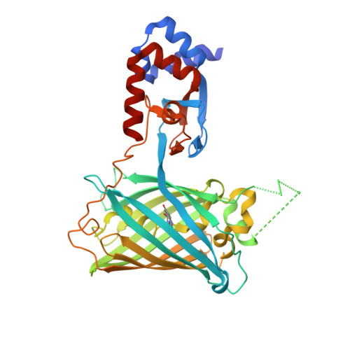
    cAMP is a key second messenger that regulates diverse cellular functions including neural plasticity. However, the spatiotemporal dynamics of intracellular cAMP in intact organisms are largely unknown due to low sensitivity and/or brightness of current genetically encoded fluorescent cAMP indicators. Here, we report the development of the new circularly permuted GFP (cpGFP)-based cAMP indicator G-Flamp1, which exhibits a large fluorescence increase (a maximum ΔF/F 0 of 1100% in HEK293T cells), decent brightness, appropriate affinity (a K d of 2.17 μM) and fast response kinetics (an association and dissociation half-time of 0.20 and 0.087 s, respectively). Furthermore, the crystal structure of the cAMP-bound G-Flamp1 reveals one linker connecting the cAMP-binding domain to cpGFP adopts a distorted β-strand conformation that may serve as a fluorescence modulation switch. We demonstrate that G-Flamp1 enables sensitive monitoring of endogenous cAMP signals in brain regions that are implicated in learning and motor control in living organisms such as fruit flies and mice.


  • Organizational Affiliation
    • Research Center for Biomedical Optics and Molecular Imaging, Shenzhen Key Laboratory for Molecular Imaging, Guangdong Provincial Key Laboratory of Biomedical Optical Imaging Technology, Shenzhen Institute of Advanced Technology, Chinese Academy of Sciences, Shenzhen, 518055, China.

Macromolecule Content 

  • Total Structure Weight: 85.9 kDa 
  • Atom Count: 5,855 
  • Modeled Residue Count: 719 
  • Deposited Residue Count: 774 
  • Unique protein chains: 1

Macromolecules

Find similar proteins by:|  3D Structure
Entity ID: 1
MoleculeChains  Sequence LengthOrganismDetailsImage
Chimera of Cyclic nucleotide-gated potassium channel mll3241 and Yellow fluorescent protein
A, B
387Mesorhizobium japonicum MAFF 303099Aequorea victoriaMutation(s): 11 
Gene Names: mll3241yfp
UniProt
Find proteins for P42212 (Aequorea victoria)
Explore P42212 
Go to UniProtKB:  P42212
Find proteins for Q98GN8 (Mesorhizobium japonicum (strain LMG 29417 / CECT 9101 / MAFF 303099))
Explore Q98GN8 
Go to UniProtKB:  Q98GN8
Entity Groups
Sequence Clusters30% Identity50% Identity70% Identity90% Identity95% Identity100% Identity
UniProt GroupsP42212Q98GN8
Sequence Annotations
Expand
Reference Sequence

Small Molecules

Modified Residues  1 Unique
IDChains TypeFormula2D DiagramParent
CRO
Query on CRO
A, B
L-PEPTIDE LINKINGC15 H17 N3 O5THR, TYR, GLY

Experimental Data & Validation

Experimental Data

  • Method: X-RAY DIFFRACTION
  • Resolution: 2.25 Å
  • R-Value Free:  0.223 (Depositor), 0.224 (DCC) 
  • R-Value Work:  0.178 (Depositor), 0.180 (DCC) 
  • R-Value Observed: 0.180 (Depositor) 
Space Group: P 21 21 21
Unit Cell:
Length ( Å )Angle ( ˚ )
a = 87.836α = 90
b = 94.688β = 90
c = 109.985γ = 90
Software Package:
Software NamePurpose
PHENIXrefinement
HKL-3000data reduction
HKL-3000data scaling
PHASERphasing

Structure Validation

View Full Validation Report


Ligand Structure Quality Assessment 


Entry History 

Deposition Data

Revision History  (Full details and data files)

  • Version 1.0: 2021-09-22
    Type: Initial release
  • Version 1.1: 2022-10-05
    Changes: Database references, Structure summary
  • Version 1.2: 2023-11-29
    Changes: Data collection, Refinement description
  • Version 1.3: 2024-11-06
    Changes: Structure summary
  • Version 2.0: 2026-03-18
    Changes: Polymer sequence