6M2V

Crystal structure of UHRF1 SRA complexed with fully-mCHG DNA.


Experimental Data Snapshot

  • Method: X-RAY DIFFRACTION
  • Resolution: 3.00 Å
  • R-Value Free: 0.276 
  • R-Value Work: 0.234 
  • R-Value Observed: 0.236 

Starting Model: experimental
View more details

wwPDB Validation   3D Report Full Report


This is version 1.2 of the entry. See complete history


Literature

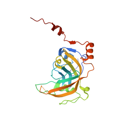
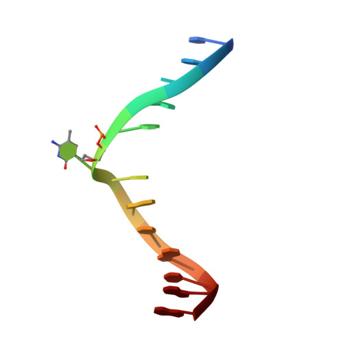
Mechanistic insights into recognition of symmetric methylated cytosines in CpG and non-CpG DNA by UHRF1 SRA.

Abhishek, S.Nakarakanti, N.K.Deeksha, W.Rajakumara, E.

(2021) Int J Biol Macromol 170: 514-522

  • DOI: https://doi.org/10.1016/j.ijbiomac.2020.12.149
  • Primary Citation of Related Structures:  
    6M2V

  • PubMed Abstract: 

    Non-CpG DNA methylation (non-mCpG) is enriched in the genome of brain neurons and germline cells in mammals. Accumulation of non-mCpG during postnatal brain development correlates with gene regulation and inactivation of distal regulatory elements. Recently, UHRF1 has been found to contribute to de novo non-CpG methylation, however, whether UHRF1 could recognize non-mCpG is unknown. Here, we have demonstrated through calorimetric measurements that the UHRF1 SRA can recognize mCpH and fully-mCpHpG, types of non-mCpG. Our ITC binding studies endorse the preferential reading of hemi-mCpG by UHRF1 SRA and also show 6-fold weaker binding for fully-mCpG than hemi-mCpG. Despite presence of symmetrical (5-methyl cytosine) 5mCs, stoichiometry of 1:1 for UHRF1 SRA binding to fully-mCpG indicates that UHRF1 SRA may not form a stable complex with fully-mCpG DNA. Contrarily, UHRF1 SRA recognizes fully-mCpHpG with a stoichiometry of 2:1 protein to DNA duplex with binding affinity higher than fully-mCpG. Our crystal structure of UHRF1 SRA bound to fully-mCpHpG DNA reveals dual flip-out mechanism of 5mC recognition. Metadynamics studies corroborates with ITC data that UHRF1 SRA could not form a stable complex with fully-mCpG DNA. Altogether, this study demonstrates that UHRF1 SRA recognizes non-mCpG DNA and exhibits contrasting mechanisms for hemi-mCpG and fully-mCpHpG DNA recognition.


  • Organizational Affiliation

    Macromolecular Structural Biology Lab, Department of Biotechnology, Indian Institute of Technology Hyderabad, Kandi, Sangareddy, Telangana 502285, India.


Macromolecules

Find similar proteins by:  (by identity cutoff)  |  3D Structure
Entity ID: 2
MoleculeChains Sequence LengthOrganismDetailsImage
E3 ubiquitin-protein ligase UHRF1C [auth B],
D [auth A]
212Mus musculusMutation(s): 0 
Gene Names: Uhrf1Np95
EC: 2.3.2.27
UniProt & NIH Common Fund Data Resources
Find proteins for Q8VDF2 (Mus musculus)
Explore Q8VDF2 
Go to UniProtKB:  Q8VDF2
IMPC:  MGI:1338889
Entity Groups  
Sequence Clusters30% Identity50% Identity70% Identity90% Identity95% Identity100% Identity
UniProt GroupQ8VDF2
Sequence Annotations
Expand
  • Reference Sequence

Find similar nucleic acids by:  Sequence   |   3D Structure  

Entity ID: 1
MoleculeChains LengthOrganismImage
DNA (5'-D(*TP*CP*AP*CP*GP*(5CM)P*TP*GP*CP*GP*TP*GP*A)-3')A [auth C],
B [auth D]
13synthetic construct
Sequence Annotations
Expand
  • Reference Sequence
Experimental Data & Validation

Experimental Data

  • Method: X-RAY DIFFRACTION
  • Resolution: 3.00 Å
  • R-Value Free: 0.276 
  • R-Value Work: 0.234 
  • R-Value Observed: 0.236 
  • Space Group: P 21 21 21
Unit Cell:
Length ( Å )Angle ( ˚ )
a = 55.897α = 90
b = 74.829β = 90
c = 141.505γ = 90
Software Package:
Software NamePurpose
XDSdata reduction
XDSdata scaling
MOLREPphasing
PHENIXrefinement

Structure Validation

View Full Validation Report


Entry History & Funding Information

Deposition Data


Funding OrganizationLocationGrant Number
Department of Biotechnology (DBT, India)India--
Science and Engineering Research Board (SERB)India--

Revision History  (Full details and data files)

  • Version 1.0: 2021-01-13
    Type: Initial release
  • Version 1.1: 2021-01-20
    Changes: Database references
  • Version 1.2: 2023-11-29
    Changes: Data collection, Database references, Refinement description