


The crystal structure of benzoate coenzyme A ligase double mutant (H333A/I334A) in complex with 2-methyl-thiazole-5 carboxylate-AMP
Li, T.L., Adhikari, K., Malek, Z.S.To be published.
Experimental Data Snapshot
Starting Model: experimental
View more details
Entity ID: 1 | |||||
|---|---|---|---|---|---|
| Molecule | Chains | Sequence Length | Organism | Details | Image |
| Benzoate-coenzyme A ligase | 524 | Rhodopseudomonas palustris | Mutation(s): 4 Gene Names: badA | 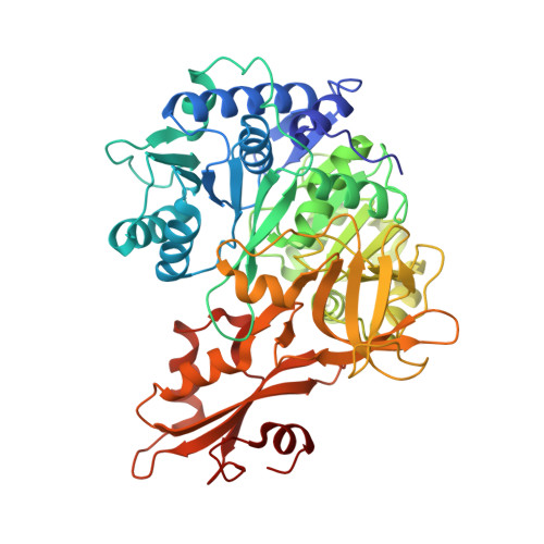 | |
UniProt | |||||
Entity Groups | |||||
| Sequence Clusters | 30% Identity50% Identity70% Identity90% Identity95% Identity100% Identity | ||||
| UniProt Group | Q93TK0 | ||||
Sequence AnnotationsExpand | |||||
Reference Sequence | |||||
| Ligands 2 Unique | |||||
|---|---|---|---|---|---|
| ID | Chains | Name / Formula / InChI Key | 2D Diagram | 3D Interactions | |
| AMP (Subject of Investigation/LOI) Download:Ideal Coordinates CCD File | E [auth A], G [auth B], I [auth C], K [auth D] | ADENOSINE MONOPHOSPHATE C10 H14 N5 O7 P UDMBCSSLTHHNCD-KQYNXXCUSA-N |  | ||
| 6V9 (Subject of Investigation/LOI) Download:Ideal Coordinates CCD File | F [auth A], H [auth B], J [auth C], L [auth D] | 2-methyl-1,3-thiazole-5-carboxylic acid C5 H5 N O2 S QCXCIYPOMMIBHO-UHFFFAOYSA-N |  | ||
| Length ( Å ) | Angle ( ˚ ) |
|---|---|
| a = 98.046 | α = 90 |
| b = 95.143 | β = 110.58 |
| c = 119.524 | γ = 90 |
| Software Name | Purpose |
|---|---|
| PHENIX | refinement |
| HKL-2000 | data reduction |
| HKL-2000 | data scaling |
| PHENIX | phasing |


