6LZI | pdb_00006lzi

Aquifex aeolicus MutL ATPase domain complexed with ADP


Experimental Data Snapshot

  • Method: X-RAY DIFFRACTION
  • Resolution: 1.69 Å
  • R-Value Free: 
    0.243 (Depositor), 0.210 (DCC) 
  • R-Value Work: 
    0.202 (Depositor), 0.172 (DCC) 
  • R-Value Observed: 
    0.205 (Depositor) 

Starting Model: experimental
View more details

wwPDB Validation 3D Report Full Report

Validation slider image for 6LZI

Ligand Structure Quality Assessment 


This is version 1.3 of the entry. See complete history

Literature

A Lynch syndrome-associated mutation at a Bergerat ATP-binding fold destabilizes the structure of the DNA mismatch repair endonuclease MutL.

Izuhara, K.Fukui, K.Murakawa, T.Baba, S.Kumasaka, T.Uchiyama, K.Yano, T.

(2020) J Biological Chem 295: 11643-11655

  • DOI: https://doi.org/10.1074/jbc.RA120.013576
  • Primary Citation Related Structures: 
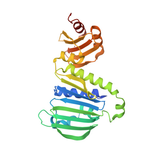
    6LZI, 6LZJ, 6LZK

  • PubMed Abstract: 

    In humans, mutations in genes encoding homologs of the DNA mismatch repair endonuclease MutL cause a hereditary cancer that is known as Lynch syndrome. Here, we determined the crystal structures of the N-terminal domain (NTD) of MutL from the thermophilic eubacterium Aquifex aeolicus (aqMutL) complexed with ATP analogs at 1.69-1.73 Å. The structures revealed significant structural similarities to those of a human MutL homolog, postmeiotic segregation increased 2 (PMS2). We introduced five Lynch syndrome-associated mutations clinically found in human PMS2 into the aqMutL NTD and investigated the protein stability, ATPase activity, and DNA-binding ability of these protein variants. Among the mutations studied, the most unexpected results were obtained for the residue Ser34. Ser34 (Ser46 in PMS2) is located at a previously identified Bergerat ATP-binding fold. We found that the S34I aqMutL NTD retains ATPase and DNA-binding activities. Interestingly, CD spectrometry and trypsin-limited proteolysis indicated the disruption of a secondary structure element of the S34I NTD, destabilizing the overall structure of the aqMutL NTD. In agreement with this, the recombinant human PMS2 S46I NTD was easily digested in the host Escherichia coli cells. Moreover, other mutations resulted in reduced DNA-binding or ATPase activity. In summary, using the thermostable aqMutL protein as a model molecule, we have experimentally determined the effects of the mutations on MutL endonuclease; we discuss the pathological effects of the corresponding mutations in human PMS2.


  • Organizational Affiliation
    • Department of Biochemistry, Osaka Medical College, Takatsuki, Osaka, Japan.

Macromolecule Content 

  • Total Structure Weight: 36.65 kDa 
  • Atom Count: 2,186 
  • Modeled Residue Count: 240 
  • Deposited Residue Count: 308 
  • Unique protein chains: 1

Macromolecules

Find similar proteins by:|  3D Structure
Entity ID: 1
MoleculeChains  Sequence LengthOrganismDetailsImage
DNA mismatch repair protein MutL308Aquifex aeolicus VF5Mutation(s): 0 
Gene Names: mutLaq_1578
UniProt
Find proteins for O67518 (Aquifex aeolicus (strain VF5))
Explore O67518 
Go to UniProtKB:  O67518
Entity Groups
Sequence Clusters30% Identity50% Identity70% Identity90% Identity95% Identity100% Identity
UniProt GroupO67518
Sequence Annotations
Expand
Reference Sequence

Small Molecules

Ligands 3 Unique
IDChains Name / Formula / InChI Key2D Diagram3D Interactions
ADP
(Subject of Investigation/LOI)

Query on ADP


Download:Ideal Coordinates CCD File
H [auth A]ADENOSINE-5'-DIPHOSPHATE
C10 H15 N5 O10 P2
XTWYTFMLZFPYCI-KQYNXXCUSA-N
PEG

Query on PEG


Download:Ideal Coordinates CCD File
C [auth A],
D [auth A],
E [auth A],
F [auth A],
G [auth A]
DI(HYDROXYETHYL)ETHER
C4 H10 O3
MTHSVFCYNBDYFN-UHFFFAOYSA-N
MG

Query on MG


Download:Ideal Coordinates CCD File
B [auth A]MAGNESIUM ION
Mg
JLVVSXFLKOJNIY-UHFFFAOYSA-N

Experimental Data & Validation

Experimental Data

  • Method: X-RAY DIFFRACTION
  • Resolution: 1.69 Å
  • R-Value Free:  0.243 (Depositor), 0.210 (DCC) 
  • R-Value Work:  0.202 (Depositor), 0.172 (DCC) 
  • R-Value Observed: 0.205 (Depositor) 
Space Group: P 1
Unit Cell:
Length ( Å )Angle ( ˚ )
a = 35.017α = 80.978
b = 42.906β = 71.521
c = 53.845γ = 77.707
Software Package:
Software NamePurpose
PHENIXrefinement
HKL-2000data processing
HKL-2000data scaling
PHENIXphasing
Cootmodel building
PHENIXmodel building
HKL-2000data reduction

Structure Validation

View Full Validation Report


Ligand Structure Quality Assessment 


Entry History 

& Funding Information

Deposition Data


Funding OrganizationLocationGrant Number
Japan Society for the Promotion of Science (JSPS)Japan19K07376

Revision History  (Full details and data files)

  • Version 1.0: 2020-07-01
    Type: Initial release
  • Version 1.1: 2020-07-08
    Changes: Database references
  • Version 1.2: 2020-09-02
    Changes: Database references
  • Version 1.3: 2023-11-29
    Changes: Data collection, Database references, Refinement description