


Structural studies reveal the molecular mechanism of isocitrate lyase from Chloroflexus aurantiacus
Lee, S.H., Kim, K.J.To be published.
Experimental Data Snapshot
Starting Model: experimental
View more details
Entity ID: 1 | |||||
|---|---|---|---|---|---|
| Molecule | Chains | Sequence Length | Organism | Details | Image |
| Isocitrate lyase | 436 | Chloroflexus aurantiacus J-10-fl | Mutation(s): 0 Gene Names: Caur_3889 EC: 4.1.3.1 | 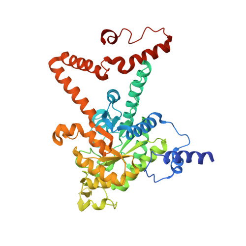 | |
UniProt | |||||
Entity Groups | |||||
| Sequence Clusters | 30% Identity50% Identity70% Identity90% Identity95% Identity100% Identity | ||||
| UniProt Group | A9WDE7 | ||||
Sequence AnnotationsExpand | |||||
Reference Sequence | |||||
| Ligands 3 Unique | |||||
|---|---|---|---|---|---|
| ID | Chains | Name / Formula / InChI Key | 2D Diagram | 3D Interactions | |
| ICT (Subject of Investigation/LOI) Download:Ideal Coordinates CCD File | BA [auth S] GA [auth V] I [auth A] L [auth D] O [auth G] | ISOCITRIC ACID C6 H8 O7 ODBLHEXUDAPZAU-ZAFYKAAXSA-N |  | ||
| GOL Download:Ideal Coordinates CCD File | CA [auth S] DA [auth S] EA [auth S] HA [auth V] IA [auth V] | GLYCEROL C3 H8 O3 PEDCQBHIVMGVHV-UHFFFAOYSA-N |  | ||
| MN (Subject of Investigation/LOI) Download:Ideal Coordinates CCD File | AA [auth P] FA [auth S] JA [auth V] K [auth A] N [auth D] | MANGANESE (II) ION Mn WAEMQWOKJMHJLA-UHFFFAOYSA-N |  | ||
| Length ( Å ) | Angle ( ˚ ) |
|---|---|
| a = 157.314 | α = 90 |
| b = 157.314 | β = 90 |
| c = 197.707 | γ = 120 |
| Software Name | Purpose |
|---|---|
| HKL-2000 | data reduction |
| HKL-2000 | data scaling |
| REFMAC | refinement |
| PDB_EXTRACT | data extraction |
| MOLREP | phasing |


