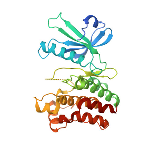
6LK6 | pdb_00006lk6

MLKL mutant - T357AS358A


Experimental Data Snapshot

  • Method: X-RAY DIFFRACTION
  • Resolution: 2.41 Å
  • R-Value Free: 
    0.247 (Depositor), 0.250 (DCC) 
  • R-Value Work: 
    0.209 (Depositor), 0.213 (DCC) 
  • R-Value Observed: 
    0.211 (Depositor) 

Starting Model: experimental
View more details

wwPDB Validation 3D Report Full Report

Validation slider image for 6LK6

This is version 1.2 of the entry. See complete history. 

Literature

The MLKL kinase-like domain dimerization is an indispensable step of mammalian MLKL activation in necroptosis signaling.

Zhang, Y., Liu, J., Yu, D., Zhu, X., Liu, X., Liao, J., Li, S., Wang, H.

(2021) Cell Death Dis 12: 638-638

  • DOI: https://doi.org/10.1038/s41419-021-03859-6
  • Primary Citation Related Structures: 
    6LK5, 6LK6

  • PubMed Abstract: 

    MLKL phosphorylation by RIP3 is the commitment step of necroptosis execution, which could induce MLKL activation featured as MLKL monomer-oligomer transition. Here, we reported that the dimerization of the MLKL kinase-like domain was the direct consequence of RIP3 triggered MLKL-phosphorylation. Two inter-dimer interfaces were found in the crystal structure of human MLKL. Mutations destroying both interfaces could prevent RIP3-induced MLKL oligomerization and necroptosis efficiently. Moreover, we confirmed MLKL self-assembly by the internal coiled-coil region is necessary for MLKL oligomerization and function. The mutations disrupting coiled-coil self-assembly repressed necroptosis, but it did not prevent RIP3-induced dimerization of the MLKL kinase-like domain. So that, MLKL activation is a sequential process, which begins with kinase-like domain dimerization, and followed by internal coiled-coil region self-assembly to form a proper MLKL oligomer. Besides human MLKL, structural and functional analysis showed the kinase-like domain dimerization was conserved among mammalian species, suggesting it is a general step of the RIP3-induced MLKL activation process.


  • Organizational Affiliation: 
    • School of Life Science and Technology, ShanghaiTech University, 393 Middle Huaxia Road, Pudong, Shanghai, 201210, China.

Macromolecule Content 

  • Total Structure Weight: 33.59 kDa 
  • Atom Count: 2,158 
  • Modeled Residue Count: 265 
  • Deposited Residue Count: 293 
  • Unique protein chains: 1

Macromolecules

Find similar proteins by:|  3D Structure
Entity ID: 1
MoleculeChains  Sequence LengthOrganismDetailsImage
Mixed lineage kinase domain-like protein293Homo sapiensMutation(s): 2 
Gene Names: MLKL
UniProt & NIH Common Fund Data Resources
Find proteins for Q8NB16 (Homo sapiens)
Explore Q8NB16 
Go to UniProtKB:  Q8NB16
PHAROS:  Q8NB16
GTEx:  ENSG00000168404 
Entity Groups
Sequence Clusters30% Identity50% Identity70% Identity90% Identity95% Identity100% Identity
UniProt GroupQ8NB16
Sequence Annotations
Expand
Reference Sequence

Experimental Data & Validation

Experimental Data

  • Method: X-RAY DIFFRACTION
  • Resolution: 2.41 Å
  • R-Value Free:  0.247 (Depositor), 0.250 (DCC) 
  • R-Value Work:  0.209 (Depositor), 0.213 (DCC) 
  • R-Value Observed: 0.211 (Depositor) 
Space Group: C 2 2 21
Unit Cell:
Length ( Å )Angle ( ˚ )
a = 70.821α = 90
b = 75.709β = 90
c = 128.494γ = 90
Software Package:
Software NamePurpose
REFMACrefinement
PDB_EXTRACTdata extraction
HKL-2000data reduction
HKL-2000data scaling
MOLREPphasing

Structure Validation

View Full Validation Report


Entry History 

& Funding Information

Deposition Data


Funding OrganizationLocationGrant Number
National Natural Science Foundation of China (NSFC)China31571427

Revision History  (Full details and data files)

  • Version 1.0: 2020-12-23
    Type: Initial release
  • Version 1.1: 2021-07-07
    Changes: Database references
  • Version 1.2: 2023-11-22
    Changes: Data collection, Database references, Refinement description