6LJI | pdb_00006lji

X-ray structure of synthetic GB1 domain with mutations K10(DVA), T11V


Experimental Data Snapshot

  • Method: X-RAY DIFFRACTION
  • Resolution: 1.84 Å
  • R-Value Free: 
    0.328 (Depositor), 0.327 (DCC) 
  • R-Value Work: 
    0.278 (Depositor), 0.281 (DCC) 
  • R-Value Observed: 
    0.281 (Depositor) 

Starting Model: experimental
View more details

wwPDB Validation 3D Report Full Report

Validation slider image for 6LJI

This is version 1.4 of the entry. See complete history

Literature

Increasing protein stability by engineering the n -> pi * interaction at the beta-turn.

Khatri, B.Majumder, P.Nagesh, J.Penmatsa, A.Chatterjee, J.

(2020) Chem Sci 11: 9480-9487

  • DOI: https://doi.org/10.1039/d0sc03060k
  • Primary Citation Related Structures: 
    6L91, 6L9B, 6L9D, 6LJI

  • PubMed Abstract: 

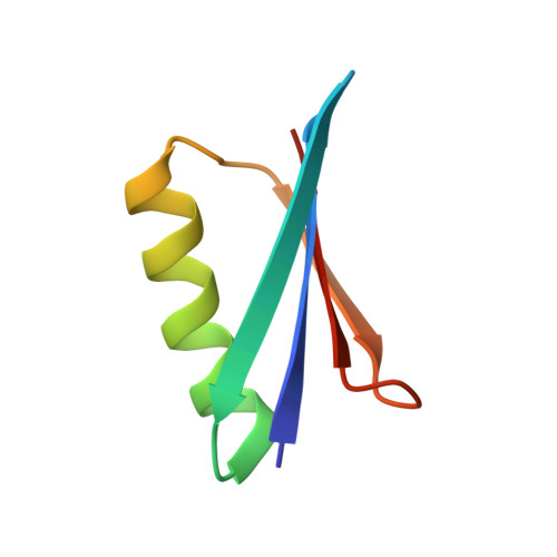
    Abundant n → π* interactions between adjacent backbone carbonyl groups, identified by statistical analysis of protein structures, are predicted to play an important role in dictating the structure of proteins. However, experimentally testing the prediction in proteins has been challenging due to the weak nature of this interaction. By amplifying the strength of the n → π* interaction via amino acid substitution and thioamide incorporation at a solvent exposed β-turn within the GB1 proteins and Pin 1 WW domain, we demonstrate that an n → π* interaction increases the structural stability of proteins by restricting the ϕ torsion angle. Our results also suggest that amino acid side-chain identity and its rotameric conformation play an important and decisive role in dictating the strength of an n → π* interaction.


  • Organizational Affiliation
    • Molecular Biophysics Unit, Indian Institute of Science Bangalore 560012 India penmatsa@iisc.ac.in jayanta@iisc.ac.in.

Macromolecule Content 

  • Total Structure Weight: 12.31 kDa 
  • Atom Count: 893 
  • Modeled Residue Count: 112 
  • Deposited Residue Count: 112 
  • Unique protein chains: 1

Macromolecules

Find similar proteins by:|  3D Structure
Entity ID: 1
MoleculeChains  Sequence LengthOrganismDetailsImage
Immunoglobulin G-binding protein G
A, B
56Streptococcus sp. 'group GMutation(s): 2 
UniProt
Find proteins for P06654 (Streptococcus sp. group G)
Explore P06654 
Go to UniProtKB:  P06654
Entity Groups
Sequence Clusters30% Identity50% Identity70% Identity90% Identity95% Identity100% Identity
UniProt GroupP06654
Sequence Annotations
Expand
Reference Sequence

Experimental Data & Validation

Experimental Data

  • Method: X-RAY DIFFRACTION
  • Resolution: 1.84 Å
  • R-Value Free:  0.328 (Depositor), 0.327 (DCC) 
  • R-Value Work:  0.278 (Depositor), 0.281 (DCC) 
  • R-Value Observed: 0.281 (Depositor) 
Space Group: C 1 2 1
Unit Cell:
Length ( Å )Angle ( ˚ )
a = 90.722α = 90
b = 23.066β = 109.04
c = 48.395γ = 90
Software Package:
Software NamePurpose
PHENIXrefinement
Aimlessdata scaling
PDB_EXTRACTdata extraction
XDSdata reduction
PHENIXphasing

Structure Validation

View Full Validation Report


Entry History 

& Funding Information

Deposition Data


Funding OrganizationLocationGrant Number
Department of Science & Technology (DST, India)IndiaEMR/2016/006193
Department of Science & Technology (DST, India)IndiaIR/SO/LU/0003/2010-PHASE-II

Revision History  (Full details and data files)

  • Version 1.0: 2020-08-12
    Type: Initial release
  • Version 1.1: 2021-08-04
    Changes: Database references
  • Version 1.2: 2022-02-09
    Changes: Database references
  • Version 1.3: 2023-11-22
    Changes: Data collection, Refinement description
  • Version 1.4: 2024-10-23
    Changes: Structure summary