6LF2 | pdb_00006lf2

SeviL bound to asialo-GM1 saccharide


Experimental Data Snapshot

  • Method: X-RAY DIFFRACTION
  • Resolution: 1.60 Å
  • R-Value Free: 
    0.228 (Depositor), 0.229 (DCC) 
  • R-Value Work: 
    0.195 (Depositor), 0.200 (DCC) 
  • R-Value Observed: 
    0.197 (Depositor) 

Starting Model: experimental
View more details

wwPDB Validation 3D Report Full Report

Validation slider image for 6LF2

This is version 1.2 of the entry. See complete history

Literature

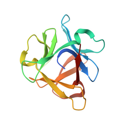
The structure of SeviL, a GM1b/asialo-GM1 binding R-type lectin from the mussel Mytilisepta virgata.

Kamata, K.Mizutani, K.Takahashi, K.Marchetti, R.Silipo, A.Addy, C.Park, S.Y.Fujii, Y.Fujita, H.Konuma, T.Ikegami, T.Ozeki, Y.Tame, J.R.H.

(2020) Sci Rep 10: 22102-22102

  • DOI: https://doi.org/10.1038/s41598-020-78926-7
  • Primary Citation Related Structures: 
    6LF1, 6LF2

  • PubMed Abstract: 

    SeviL is a recently isolated lectin found to bind to the linear saccharides of the ganglioside GM1b (Neu5Ac[Formula: see text](2-3)Gal[Formula: see text](1-3)GalNAc[Formula: see text](1-4)Gal[Formula: see text](1-4)Glc) and its precursor, asialo-GM1 (Gal[Formula: see text](1-3)GalNAc[Formula: see text](1-4)Gal[Formula: see text](1-4)Glc). The crystal structures of recombinant SeviL have been determined in the presence and absence of ligand. The protein belongs to the [Formula: see text]-trefoil family, but shows only weak sequence similarity to known structures. SeviL forms a dimer in solution, with one binding site per subunit, close to the subunit interface. Molecular details of glycan recognition by SeviL in solution were analysed by ligand- and protein-based NMR techniques as well as ligand binding assays. SeviL shows no interaction with GM1 due to steric hindrance with the sialic acid branch that is absent from GM1b. This unusual specificity makes SeviL of great interest for the detection and control of certain cancer cells, and cells of the immune system, that display asialo-GM1.


  • Organizational Affiliation
    • Graduate School of Medical Life Science, Yokohama City University, 1-7-29 Suehiro, Yokohama, Kanagawa, 230-0045, Japan.

Macromolecule Content 

  • Total Structure Weight: 32.11 kDa 
  • Atom Count: 2,357 
  • Modeled Residue Count: 256 
  • Deposited Residue Count: 264 
  • Unique protein chains: 1

Macromolecules

Find similar proteins by:|  3D Structure
Entity ID: 1
MoleculeChains  Sequence LengthOrganismDetailsImage
SeviL
A, B
132Mytilisepta virgataMutation(s): 0 
UniProt
Find proteins for A0A646QV53 (Mytilisepta virgata)
Explore A0A646QV53 
Go to UniProtKB:  A0A646QV53
Entity Groups
Sequence Clusters30% Identity50% Identity70% Identity90% Identity95% Identity100% Identity
UniProt GroupA0A646QV53
Sequence Annotations
Expand
Reference Sequence

Oligosaccharides

Help  
Entity ID: 2
MoleculeChains Length2D Diagram GlycosylationD Interactions
beta-D-galactopyranose-(1-3)-2-acetamido-2-deoxy-beta-D-galactopyranose-(1-4)-beta-D-galactopyranose-(1-4)-beta-D-glucopyranose
C, D
4N/A
Glycosylation Resources
GlyTouCan: G21856LC
GlyCosmos: G21856LC
GlyGen: G21856LC

Experimental Data & Validation

Experimental Data

  • Method: X-RAY DIFFRACTION
  • Resolution: 1.60 Å
  • R-Value Free:  0.228 (Depositor), 0.229 (DCC) 
  • R-Value Work:  0.195 (Depositor), 0.200 (DCC) 
  • R-Value Observed: 0.197 (Depositor) 
Space Group: P 41 21 2
Unit Cell:
Length ( Å )Angle ( ˚ )
a = 61.59α = 90
b = 61.59β = 90
c = 136.783γ = 90
Software Package:
Software NamePurpose
PHENIXrefinement
REFMACrefinement
HKL-2000data reduction
SCALEPACKdata scaling
MOLREPphasing

Structure Validation

View Full Validation Report


Entry History 

& Funding Information

Deposition Data


Funding OrganizationLocationGrant Number
Japan Science and TechnologyJapanJP18H02413

Revision History  (Full details and data files)

  • Version 1.0: 2020-12-02
    Type: Initial release
  • Version 1.1: 2020-12-30
    Changes: Database references
  • Version 1.2: 2023-11-22
    Changes: Data collection, Database references, Refinement description