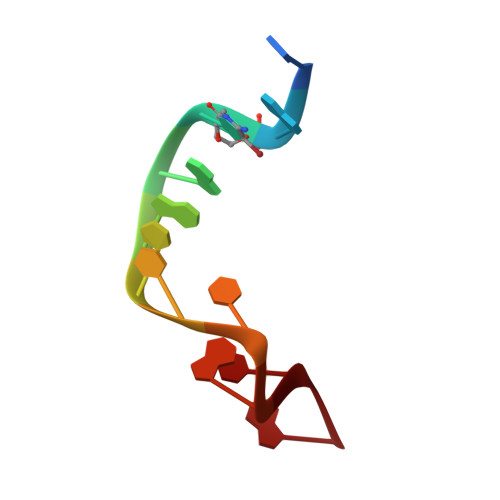
6LBW | pdb_00006lbw

Crystal structure of Ag-mediated base pairs in uncanonical DNA duplex


Experimental Data Snapshot

  • Method: X-RAY DIFFRACTION
  • Resolution: 1.50 Å
  • R-Value Free: 
    0.243 (Depositor), 0.245 (DCC) 
  • R-Value Work: 
    0.214 (Depositor), 0.217 (DCC) 
  • R-Value Observed: 
    0.217 (Depositor) 

wwPDB Validation 3D Report Full Report

Validation slider image for 6LBW

This is version 1.4 of the entry. See complete history

Literature

Crystallographic Structure of Novel Types of Ag I -Mediated Base Pairs in Non-canonical DNA Duplex Containing 2'-O,4'-C-Methylene Bridged Nucleic Acids.

Nakagawa, O.Aoyama, H.Fujii, A.Kishimoto, Y.Obika, S.

(2021) Chemistry 27: 3842-3848

  • DOI: https://doi.org/10.1002/chem.202004819
  • Primary Citation Related Structures: 
    6LBW

  • PubMed Abstract: 

    Metal-mediated base pairs have widespread applications, such as in DNA-metal nanodevices and sensors. Here, we focused on their sugar conformation in duplexes and observed the crystallographic structure of the non-canonical DNA/DNA duplex containing 2'-O,4'-C-methylene bridged nucleic acid in the presence of Ag I ions. The X-ray crystallographic structure was successfully obtained at a resolution of 1.5 Å. A novel type of Ag I -mediated base pair between the N1 positions of anti-conformation of adenines in the duplex was observed. In the central non-canonical region, a hexad nucleobase structure containing Ag I -mediated base pairs between the N7 positions of guanines was formed. A highly bent non-canonical structure was formed at the origin of Ag I -mediated base pairs in the central region. The bent duplex structure induced by the addition of Ag I ions might become a powerful tool for dynamic structural changes in DNA nanotechnology applications.


  • Organizational Affiliation
    • Graduate School of Pharmaceutical Sciences, Osaka University, 1-6 Yamadaoka Suita, Osaka, 565-0871, Japan.

Macromolecule Content 

  • Total Structure Weight: 7.13 kDa 
  • Atom Count: 510 
  • Modeled Residue Count: 22 
  • Deposited Residue Count: 22 
  • Unique nucleic acid chains: 1

Macromolecules

Find similar nucleic acids by:  Sequence
Entity ID: 1
MoleculeChains LengthOrganismImage
DNA (5'-D(*CP*GP*(CBR)P*GP*AP*(LCC)P*TP*CP*GP*CP*G)-3')
A, B
11synthetic construct
Sequence Annotations
Expand
Reference Sequence

Experimental Data & Validation

Experimental Data

  • Method: X-RAY DIFFRACTION
  • Resolution: 1.50 Å
  • R-Value Free:  0.243 (Depositor), 0.245 (DCC) 
  • R-Value Work:  0.214 (Depositor), 0.217 (DCC) 
  • R-Value Observed: 0.217 (Depositor) 
Space Group: F 2 2 2
Unit Cell:
Length ( Å )Angle ( ˚ )
a = 28.56α = 90
b = 68.11β = 90
c = 118.85γ = 90
Software Package:
Software NamePurpose
PHENIXrefinement
XDSdata reduction
XSCALEdata scaling
PDB_EXTRACTdata extraction
SHELXDEphasing

Structure Validation

View Full Validation Report


Entry History 

Deposition Data

Revision History  (Full details and data files)

  • Version 1.0: 2020-11-18
    Type: Initial release
  • Version 1.1: 2020-12-16
    Changes: Database references, Structure summary
  • Version 1.2: 2020-12-23
    Changes: Database references
  • Version 1.3: 2021-03-03
    Changes: Database references
  • Version 1.4: 2024-03-27
    Changes: Data collection, Database references