6L9C | pdb_00006l9c

Neutron structure of copper amine oxidase from Arthrobacter glibiformis at pD 7.4


Experimental Data Snapshot

  • Method: X-RAY DIFFRACTION
  • Resolution: 1.14 Å
  • R-Value Free: 
    0.181 (Depositor), 0.181 (DCC) 
  • R-Value Work: 
    0.168 (Depositor), 0.168 (DCC) 
  • R-Value Observed: 
    0.168 (Depositor) 

  • Method: NEUTRON DIFFRACTION
  • Resolution: 1.72 Å
  • R-Value Free: 
    0.209 (Depositor) 
  • R-Value Work: 
    0.180 (Depositor) 
  • R-Value Observed: 
    0.181 (Depositor) 

Starting Model: experimental
View more details

wwPDB Validation 3D Report Full Report

Validation slider image for 6L9C

This is version 1.3 of the entry. See complete history

Literature

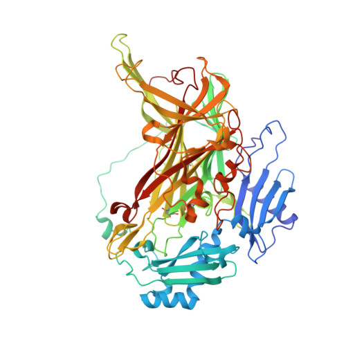
Neutron crystallography of copper amine oxidase reveals keto/enolate interconversion of the quinone cofactor and unusual proton sharing.

Murakawa, T.Kurihara, K.Shoji, M.Shibazaki, C.Sunami, T.Tamada, T.Yano, N.Yamada, T.Kusaka, K.Suzuki, M.Shigeta, Y.Kuroki, R.Hayashi, H.Yano, T.Tanizawa, K.Adachi, M.Okajima, T.

(2020) Proc Natl Acad Sci U S A 117: 10818-10824

  • DOI: https://doi.org/10.1073/pnas.1922538117
  • Primary Citation Related Structures: 
    6L9C

  • PubMed Abstract: 

    Recent advances in neutron crystallographic studies have provided structural bases for quantum behaviors of protons observed in enzymatic reactions. Thus, we resolved the neutron crystal structure of a bacterial copper (Cu) amine oxidase (CAO), which contains a prosthetic Cu ion and a protein-derived redox cofactor, topa quinone (TPQ). We solved hitherto unknown structures of the active site, including a keto/enolate equilibrium of the cofactor with a nonplanar quinone ring, unusual proton sharing between the cofactor and the catalytic base, and metal-induced deprotonation of a histidine residue that coordinates to the Cu. Our findings show a refined active-site structure that gives detailed information on the protonation state of dissociable groups, such as the quinone cofactor, which are critical for catalytic reactions.


  • Organizational Affiliation
    • Department of Biochemistry, Osaka Medical College, 569-8686 Takatsuki, Osaka, Japan; murakawa@osaka-med.ac.jp tokajima@sanken.osaka-u.ac.jp.

Macromolecule Content 

  • Total Structure Weight: 69.08 kDa 
  • Atom Count: 6,258 
  • Modeled Residue Count: 622 
  • Deposited Residue Count: 622 
  • Unique protein chains: 1

Macromolecules

Find similar proteins by:|  3D Structure
Entity ID: 1
MoleculeChains  Sequence LengthOrganismDetailsImage
Phenylethylamine oxidaseA [auth X]621Arthrobacter globiformisMutation(s): 0 
EC: 1.4.3.21
UniProt
Find proteins for P46881 (Arthrobacter globiformis)
Explore P46881 
Go to UniProtKB:  P46881
Entity Groups
Sequence Clusters30% Identity50% Identity70% Identity90% Identity95% Identity100% Identity
UniProt GroupP46881
Sequence Annotations
Expand
Reference Sequence

Small Molecules

Ligands 2 Unique
IDChains Name / Formula / InChI Key2D Diagram3D Interactions
CU

Query on CU


Download:Ideal Coordinates CCD File
B [auth X]COPPER (II) ION
Cu
JPVYNHNXODAKFH-UHFFFAOYSA-N
NA

Query on NA


Download:Ideal Coordinates CCD File
C [auth X]SODIUM ION
Na
FKNQFGJONOIPTF-UHFFFAOYSA-N
Modified Residues  3 Unique
IDChains TypeFormula2D DiagramParent
ASA
Query on ASA
A [auth X]L-PEPTIDE LINKINGC4 H7 N O3ASP
E9C
Query on E9C
A [auth X]L-PEPTIDE LINKINGC9 H9 N O5TYR
TPQ
Query on TPQ
A [auth X]L-PEPTIDE LINKINGC9 H9 N O5TYR

Experimental Data & Validation

Experimental Data

  • Method: X-RAY DIFFRACTION
  • Resolution: 1.14 Å
  • R-Value Free:  0.181 (Depositor), 0.181 (DCC) 
  • R-Value Work:  0.168 (Depositor), 0.168 (DCC) 
  • R-Value Observed: 0.168 (Depositor) 
  • Method: NEUTRON DIFFRACTION
  • Resolution: 1.72 Å
  • R-Value Free:  0.209 (Depositor) 
  • R-Value Work:  0.180 (Depositor) 
  • R-Value Observed: 0.181 (Depositor) 
Space Group: C 1 2 1
Unit Cell:
Length ( Å )Angle ( ˚ )
a = 157.548α = 90
b = 61.779β = 112.13
c = 92.332γ = 90
Software Package:
Software NamePurpose
PHENIXrefinement
HKL-2000data scaling
PDB_EXTRACTdata extraction
HKL-2000data reduction
PHASERphasing

Structure Validation

View Full Validation Report


Entry History 

& Funding Information

Deposition Data


Funding OrganizationLocationGrant Number
Japan Society for the Promotion of ScienceJapanJP23770127, JP26440037, JP15K05573, JP16KT0055, 19K05694

Revision History  (Full details and data files)

  • Version 1.0: 2020-04-29
    Type: Initial release
  • Version 1.1: 2020-05-20
    Changes: Database references
  • Version 1.2: 2020-06-03
    Changes: Database references
  • Version 1.3: 2023-11-22
    Changes: Data collection, Database references, Derived calculations, Refinement description