6L8F | pdb_00006l8f

Crystal structure of heterotetrameric complex of YoeB-YefM toxin-antitoxin from Staphylococcus aureus.


Experimental Data Snapshot

  • Method: X-RAY DIFFRACTION
  • Resolution: 2.40 Å
  • R-Value Free: 
    0.223 (Depositor), 0.227 (DCC) 
  • R-Value Work: 
    0.190 (Depositor), 0.193 (DCC) 
  • R-Value Observed: 
    0.191 (Depositor) 

Starting Model: experimental
View more details

wwPDB Validation 3D Report Full Report

Validation slider image for 6L8F

This is version 1.3 of the entry. See complete history

Literature

Distinct oligomeric structures of the YoeB-YefM complex provide insights into the conditional cooperativity of type II toxin-antitoxin system.

Xue, L.Yue, J.Ke, J.Khan, M.H.Wen, W.Sun, B.Zhu, Z.Niu, L.

(2020) Nucleic Acids Res 48: 10527-10541

  • DOI: https://doi.org/10.1093/nar/gkaa706
  • Primary Citation Related Structures: 
    6L8E, 6L8F, 7CUA

  • PubMed Abstract: 

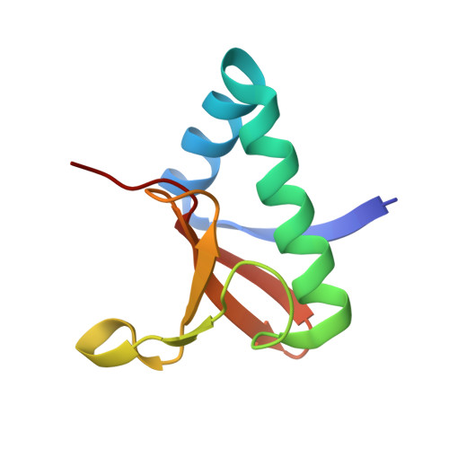
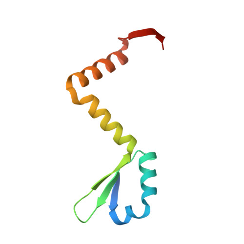
    YoeB-YefM, the widespread type II toxin-antitoxin (TA) module, binds to its own promoter to autoregulate its transcription: repress or induce transcription under normal or stress conditions, respectively. It remains unclear how YoeB-YefM regulates its transcription depending on the YoeB to YefM TA ratio. We find that YoeB-YefM complex from S.aureus exists as two distinct oligomeric assemblies: heterotetramer (YoeB-YefM2-YoeB) and heterohexamer (YoeB-YefM2-YefM2-YoeB) with low and high DNA-binding affinities, respectively. Structures of the heterotetramer alone and heterohexamer bound to promoter DNA reveals that YefM C-terminal domain undergoes disorder to order transition upon YoeB binding, which allosterically affects the conformation of N-terminal DNA-binding domain. At TA ratio of 1:2, unsaturated binding of YoeB to the C-terminal regions of YefM dimer forms an optimal heterohexamer for DNA binding, and two YefM dimers with N-terminal domains dock into the adjacent major grooves of DNA to specifically recognize the 5'-TTGTACAN6AGTACAA-3' palindromic sequence, resulting in transcriptional repression. In contrast, at TA ratio of 1:1, binding of two additional YoeB molecules onto the heterohexamer induces the completely ordered conformation of YefM and disassembles the heterohexamer into two heterotetramers, which are unable to bind the promoter DNA optimally due to steric clashes, hence derepresses TA operon transcription.


  • Organizational Affiliation
    • Hefei National Laboratory for Physical Sciences at the Microscale, Division of Molecular and Cellular Biophysics, University of Science and Technology of China, Hefei, Anhui 230026, China.

Macromolecule Content 

  • Total Structure Weight: 82.92 kDa 
  • Atom Count: 5,674 
  • Modeled Residue Count: 682 
  • Deposited Residue Count: 708 
  • Unique protein chains: 2

Macromolecules

Find similar proteins by:|  3D Structure
Entity ID: 1
MoleculeChains  Sequence LengthOrganismDetailsImage
YoeBA [auth D],
C,
E [auth H],
G
88Staphylococcus aureus subsp. aureus NCTC 8325Mutation(s): 0 
Gene Names: SAOUHSC_02691
UniProt
Find proteins for Q2G286 (Staphylococcus aureus (strain NCTC 8325 / PS 47))
Explore Q2G286 
Go to UniProtKB:  Q2G286
Entity Groups
Sequence Clusters30% Identity50% Identity70% Identity90% Identity95% Identity100% Identity
UniProt GroupQ2G286
Sequence Annotations
Expand
Reference Sequence
Find similar proteins by:|  3D Structure
Entity ID: 2
MoleculeChains  Sequence LengthOrganismDetailsImage
AntitoxinB,
D [auth A],
F,
H [auth E]
89Staphylococcus aureus subsp. aureus NCTC 8325Mutation(s): 0 
Gene Names: SAOUHSC_02692
UniProt
Find proteins for Q2G285 (Staphylococcus aureus (strain NCTC 8325 / PS 47))
Explore Q2G285 
Go to UniProtKB:  Q2G285
Entity Groups
Sequence Clusters30% Identity50% Identity70% Identity90% Identity95% Identity100% Identity
UniProt GroupQ2G285
Sequence Annotations
Expand
Reference Sequence

Experimental Data & Validation

Experimental Data

  • Method: X-RAY DIFFRACTION
  • Resolution: 2.40 Å
  • R-Value Free:  0.223 (Depositor), 0.227 (DCC) 
  • R-Value Work:  0.190 (Depositor), 0.193 (DCC) 
  • R-Value Observed: 0.191 (Depositor) 
Space Group: P 61
Unit Cell:
Length ( Å )Angle ( ˚ )
a = 104.438α = 90
b = 104.438β = 90
c = 172.586γ = 120
Software Package:
Software NamePurpose
PHENIXrefinement
XDSdata reduction
Aimlessdata scaling
PHENIXphasing

Structure Validation

View Full Validation Report


Entry History 

& Funding Information

Deposition Data


Funding OrganizationLocationGrant Number
National Natural Science Foundation of China (NSFC)ChinaU1632124 to L.N.

Revision History  (Full details and data files)

  • Version 1.0: 2020-09-02
    Type: Initial release
  • Version 1.1: 2020-09-09
    Changes: Database references
  • Version 1.2: 2020-10-21
    Changes: Database references
  • Version 1.3: 2023-11-22
    Changes: Data collection, Database references, Refinement description