6L42 | pdb_00006l42

Structure of severe fever with thrombocytopenia syndrome virus L protein


Experimental Data Snapshot

  • Method: ELECTRON MICROSCOPY
  • Resolution: 3.40 Å
  • Aggregation State: PARTICLE 
  • Reconstruction Method: SINGLE PARTICLE 

wwPDB Validation 3D Report Full Report

Validation slider image for 6L42

This is version 2.2 of the entry. See complete history

Literature

Structure of severe fever with thrombocytopenia syndrome virus L protein elucidates the mechanisms of viral transcription initiation.

Wang, P.Liu, L.Liu, A.Yan, L.He, Y.Shen, S.Hu, M.Guo, Y.Liu, H.Liu, C.Lu, Y.Wang, P.Deng, F.Rao, Z.Lou, Z.

(2020) Nat Microbiol 5: 864-871

  • DOI: https://doi.org/10.1038/s41564-020-0712-2
  • Primary Citation Related Structures: 
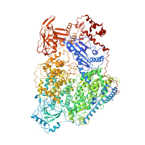
    6L42

  • PubMed Abstract: 

    Segmented negative-sense RNA viruses (sNSRVs) encode a single-polypeptide polymerase (L protein) or a heterotrimeric polymerase complex to cannibalize host messenger RNA cap structures serving as primers of transcription, and catalyse RNA synthesis. Here, we report the full-length structure of the severe fever with thrombocytopaenia syndrome virus (SFTSV) L protein, as determined by cryogenic electron microscopy at 3.4 Å, leading to an atomic model harbouring three functional parts (an endonuclease, an RNA-dependent RNA polymerase and a cap-binding domain) and two structural domains (an arm domain with a blocker motif and a carboxy-terminal lariat domain). The SFTSV L protein has a compact architecture in which its cap-binding pocket is surprisingly occupied by an Arg finger of the blocker motif, and the endonuclease active centre faces back towards the cap-binding pocket, suggesting that domain rearrangements are necessary to acquire the pre-initiation state of the active site. Our results provide insight into the complete architecture of sNSRV-encoded L protein and further the understanding of sNSRV transcription initiation.


  • Organizational Affiliation
    • MOE Key Laboratory of Protein Science and Collaborative Innovation Center of Biotherapy, School of Medicine, Tsinghua University, Beijing, China.

Macromolecule Content 

  • Total Structure Weight: 238.84 kDa 
  • Atom Count: 14,827 
  • Modeled Residue Count: 1,864 
  • Deposited Residue Count: 2,109 
  • Unique protein chains: 1

Macromolecules

Find similar proteins by:|  3D Structure
Entity ID: 1
MoleculeChains  Sequence LengthOrganismDetailsImage
RNA polymerase2,109Phlebovirus WCH/97/HN/China/2011Mutation(s): 1 
EC: 3.1 (UniProt), 2.7.7.48 (UniProt)
UniProt
Find proteins for I0DF35 (Dabie bandavirus)
Explore I0DF35 
Go to UniProtKB:  I0DF35
Entity Groups
Sequence Clusters30% Identity50% Identity70% Identity90% Identity95% Identity100% Identity
UniProt GroupI0DF35
Sequence Annotations
Expand
Reference Sequence

Small Molecules

Ligands 1 Unique
IDChains Name / Formula / InChI Key2D Diagram3D Interactions
MG

Query on MG


Download:Ideal Coordinates CCD File
B [auth A]MAGNESIUM ION
Mg
JLVVSXFLKOJNIY-UHFFFAOYSA-N

Experimental Data & Validation

Experimental Data

  • Method: ELECTRON MICROSCOPY
  • Resolution: 3.40 Å
  • Aggregation State: PARTICLE 
  • Reconstruction Method: SINGLE PARTICLE 
EM Software:
TaskSoftware PackageVersion
MODEL REFINEMENTPHENIX

Structure Validation

View Full Validation Report


Entry History 

& Funding Information

Deposition Data


Funding OrganizationLocationGrant Number
National Natural Science Foundation of China (NSFC)China--

Revision History  (Full details and data files)

  • Version 1.0: 2020-05-13
    Type: Initial release
  • Version 2.0: 2021-12-01
    Type: Coordinate replacement
    Reason: Atomic clashes
    Changes: Advisory, Atomic model, Data collection, Database references, Derived calculations, Polymer sequence, Structure summary
  • Version 2.1: 2024-05-29
    Changes: Data collection
  • Version 2.2: 2025-07-02
    Changes: Data collection, Structure summary