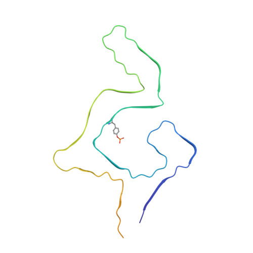
Parkinson's disease-related phosphorylation at Tyr39 rearranges alpha-synuclein amyloid fibril structure revealed by cryo-EM.
Zhao, K., Lim, Y.J., Liu, Z., Long, H., Sun, Y., Hu, J.J., Zhao, C., Tao, Y., Zhang, X., Li, D., Li, Y.M., Liu, C.(2020) Proc Natl Acad Sci U S A 117: 20305-20315
- PubMed: 32737160
- DOI: https://doi.org/10.1073/pnas.1922741117
- Primary Citation of Related Structures:
6L1T, 6L1U - PubMed Abstract:
Posttranslational modifications (PTMs) of α-synuclein (α-syn), e.g., phosphorylation, play an important role in modulating α-syn pathology in Parkinson's disease (PD) and α-synucleinopathies. Accumulation of phosphorylated α-syn fibrils in Lewy bodies and Lewy neurites is the histological hallmark of these diseases. However, it is unclear how phosphorylation relates to α-syn pathology. Here, by combining chemical synthesis and bacterial expression, we obtained homogeneous α-syn fibrils with site-specific phosphorylation at Y39, which exhibits enhanced neuronal pathology in rat primary cortical neurons. We determined the cryo-electron microscopy (cryo-EM) structure of the pY39 α-syn fibril, which reveals a fold of α-syn with pY39 in the center of the fibril core forming an electrostatic interaction network with eight charged residues in the N-terminal region of α-syn. This structure composed of residues 1 to 100 represents the largest α-syn fibril core determined so far. This work provides structural understanding on the pathology of the pY39 α-syn fibril and highlights the importance of PTMs in defining the polymorphism and pathology of amyloid fibrils in neurodegenerative diseases.
- Interdisciplinary Research Center on Biology and Chemistry, Shanghai Institute of Organic Chemistry, Chinese Academy of Sciences, Shanghai 201210, China.
Organizational Affiliation:


