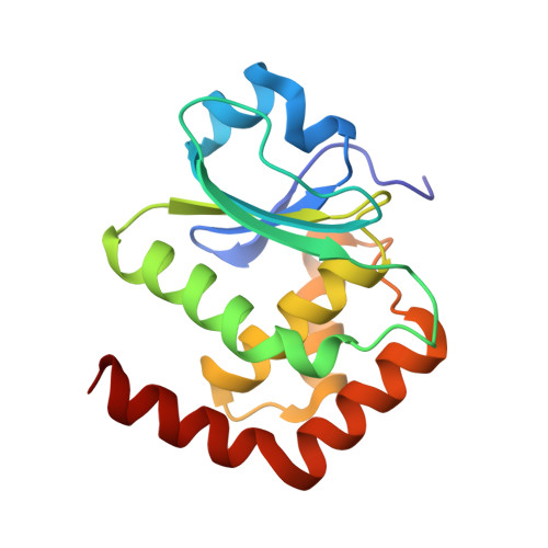
Structural Insights into the Active Site Formation of DUSP22 in N-loop-containing Protein Tyrosine Phosphatases.
Lai, C.H., Chang, C.C., Chuang, H.C., Tan, T.H., Lyu, P.C.(2020) Int J Mol Sci 21
- PubMed: 33053837
- DOI: https://doi.org/10.3390/ijms21207515
- Primary Citation of Related Structures:
6L1S, 6LMY, 6LOT, 6LOU, 6LVQ, 7C8S - PubMed Abstract:
Cysteine-based protein tyrosine phosphatases (Cys-based PTPs) perform dephosphorylation to regulate signaling pathways in cellular responses. The hydrogen bonding network in their active site plays an important conformational role and supports the phosphatase activity. Nearly half of dual-specificity phosphatases (DUSPs) use three conserved residues, including aspartate in the D-loop, serine in the P-loop, and asparagine in the N-loop, to form the hydrogen bonding network, the D-, P-, N-triloop interaction (DPN-triloop interaction). In this study, DUSP22 is used to investigate the importance of the DPN-triloop interaction in active site formation. Alanine mutations and somatic mutations of the conserved residues, D57, S93, and N128 substantially decrease catalytic efficiency ( k cat / K M ) by more than 10 2 -fold. Structural studies by NMR and crystallography reveal that each residue can perturb the three loops and induce conformational changes, indicating that the hydrogen bonding network aligns the residues in the correct positions for substrate interaction and catalysis. Studying the DPN-triloop interaction reveals the mechanism maintaining phosphatase activity in N-loop-containing PTPs and provides a foundation for further investigation of active site formation in different members of this protein class.
- Institute of Bioinformatics and Structural Biology, Department of Life Science, National Tsing Hua University, Hsinchu 30013, Taiwan.
Organizational Affiliation:


