6L0G | pdb_00006l0g

Crystal structure of dihydroorotase in complex with malate at pH6 from Saccharomyces cerevisiae


Experimental Data Snapshot

  • Method: X-RAY DIFFRACTION
  • Resolution: 2.05 Å
  • R-Value Free: 
    0.232 (Depositor), 0.232 (DCC) 
  • R-Value Work: 
    0.186 (Depositor) 
  • R-Value Observed: 
    0.189 (Depositor) 

Starting Model: experimental
View more details

wwPDB Validation 3D Report Full Report

Validation slider image for 6L0G

Ligand Structure Quality Assessment 


This is version 1.2 of the entry. See complete history

Literature

Structural basis for the interaction modes of dihydroorotase with the anticancer drugs 5-fluorouracil and 5-aminouracil.

Guan, H.H.Huang, Y.H.Lin, E.S.Chen, C.J.Huang, C.Y.

(2021) Biochem Biophys Res Commun 551: 33-37

  • DOI: https://doi.org/10.1016/j.bbrc.2021.03.001
  • Primary Citation Related Structures: 
    6L0B, 6L0F, 6L0G, 6L0H, 6L0I, 6L0K

  • PubMed Abstract: 

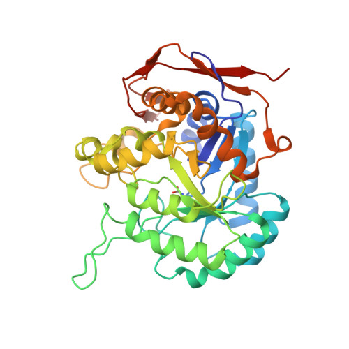
    Dihydroorotase (DHOase) is the third enzyme in the de novo biosynthesis pathway of pyrimidine nucleotides and considered an attractive target for potential antimalarial, anticancer, and antipathogen chemotherapy. Whether the FDA-approved clinical drug 5-fluorouracil (5-FU) that is used to target the enzyme thymidylate synthase for anticancer therapy can also bind to DHOase remains unknown. Here, we report the crystal structures of DHOase from Saccharomyces cerevisiae (ScDHOase) complexed with malate, 5-FU, and 5-aminouracil (5-AU). ScDHOase shares structural similarity with Escherichia coli DHOase. We also characterized the binding of 5-FU and 5-AU to ScDHOase by using the fluorescence quenching method. These complexed structures revealed that residues Arg18, Asn43, Thr106, and Ala275 of ScDHOase were involved in the 5-FU (PDB entry 6L0B) and 5-AU binding (PDB entry 6L0F). Overall, these results provide structural insights that may facilitate the development of new inhibitors targeting DHOase and constitute the 5-FU and 5-AU interactomes for further clinical chemotherapies.


  • Organizational Affiliation
    • Life Science Group, Scientific Research Division, National Synchrotron Radiation Research Center, Hsinchu, Taiwan.

Macromolecule Content 

  • Total Structure Weight: 166.97 kDa 
  • Atom Count: 12,641 
  • Modeled Residue Count: 1,455 
  • Deposited Residue Count: 1,488 
  • Unique protein chains: 1

Macromolecules

Find similar proteins by:|  3D Structure
Entity ID: 1
MoleculeChains  Sequence LengthOrganismDetailsImage
Dihydroorotase
A, B, C, D
372Saccharomyces cerevisiae S288CMutation(s): 0 
Gene Names: URA4YLR420WL9931.1
EC: 3.5.2.3
UniProt
Find proteins for P20051 (Saccharomyces cerevisiae (strain ATCC 204508 / S288c))
Explore P20051 
Go to UniProtKB:  P20051
Entity Groups
Sequence Clusters30% Identity50% Identity70% Identity90% Identity95% Identity100% Identity
UniProt GroupP20051
Sequence Annotations
Expand
Reference Sequence

Small Molecules

Ligands 2 Unique
IDChains Name / Formula / InChI Key2D Diagram3D Interactions
LMR
(Subject of Investigation/LOI)

Query on LMR


Download:Ideal Coordinates CCD File
G [auth A],
J [auth B],
M [auth C],
P [auth D]
(2S)-2-hydroxybutanedioic acid
C4 H6 O5
BJEPYKJPYRNKOW-REOHCLBHSA-N
ZN
(Subject of Investigation/LOI)

Query on ZN


Download:Ideal Coordinates CCD File
E [auth A]
F [auth A]
H [auth B]
I [auth B]
K [auth C]
E [auth A],
F [auth A],
H [auth B],
I [auth B],
K [auth C],
L [auth C],
N [auth D],
O [auth D]
ZINC ION
Zn
PTFCDOFLOPIGGS-UHFFFAOYSA-N
Modified Residues  1 Unique
IDChains TypeFormula2D DiagramParent
KCX
Query on KCX
A, B, C, D
L-PEPTIDE LINKINGC7 H14 N2 O4LYS

Experimental Data & Validation

Experimental Data

  • Method: X-RAY DIFFRACTION
  • Resolution: 2.05 Å
  • R-Value Free:  0.232 (Depositor), 0.232 (DCC) 
  • R-Value Work:  0.186 (Depositor) 
  • R-Value Observed: 0.189 (Depositor) 
Space Group: P 1 21 1
Unit Cell:
Length ( Å )Angle ( ˚ )
a = 85.44α = 90
b = 88.544β = 95.39
c = 103.526γ = 90
Software Package:
Software NamePurpose
PHENIXrefinement
PDB_EXTRACTdata extraction
HKL-2000data reduction
HKL-2000data scaling
MOLREPphasing

Structure Validation

View Full Validation Report


Ligand Structure Quality Assessment 


Entry History 

& Funding Information

Deposition Data


Funding OrganizationLocationGrant Number
Ministry of Science and Technology (Taiwan)Taiwan--

Revision History  (Full details and data files)

  • Version 1.0: 2020-12-02
    Type: Initial release
  • Version 1.1: 2021-06-16
    Changes: Database references
  • Version 1.2: 2023-11-22
    Changes: Data collection, Database references, Refinement description