6L06 | pdb_00006l06

Crystal structure of Escherichia coli phosphatidylserine decarboxylase (apo-form)


Experimental Data Snapshot

  • Method: X-RAY DIFFRACTION
  • Resolution: 2.60 Å
  • R-Value Free: 
    0.276 (Depositor), 0.278 (DCC) 
  • R-Value Work: 
    0.231 (Depositor), 0.235 (DCC) 
  • R-Value Observed: 
    0.233 (Depositor) 

wwPDB Validation 3D Report Full Report

Validation slider image for 6L06

This is version 2.1 of the entry. See complete history

Literature

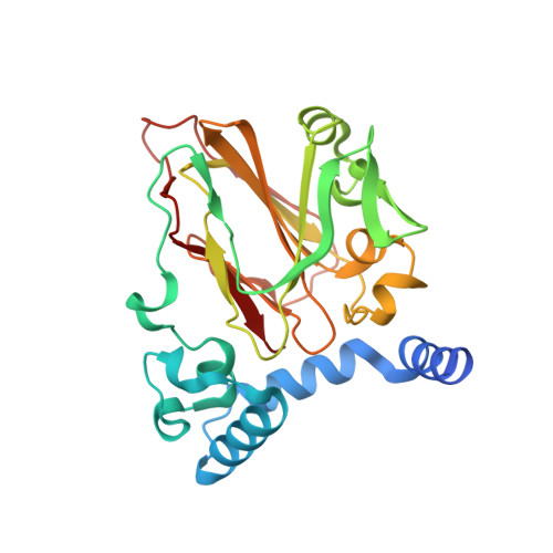
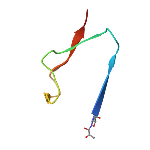
Structural Basis for Phosphatidylethanolamine Biosynthesis by Bacterial Phosphatidylserine Decarboxylase.

Watanabe, Y.Watanabe, Y.Watanabe, S.

(2020) Structure 28: 799

  • DOI: https://doi.org/10.1016/j.str.2020.04.006
  • Primary Citation Related Structures: 
    6L06, 6L07

  • PubMed Abstract: 

    In both prokaryotes and eukaryotes, phosphatidylethanolamine (PE), one of the most abundant membrane phospholipids, plays important roles in various membrane functions and is synthesized through the decarboxylation of phosphatidylserine (PS) by PS decarboxylases (PSDs). However, the catalysis and substrate recognition mechanisms of PSDs remain unclear. In this study, we focused on the PSD from Escherichia coli (EcPsd) and determined the crystal structures of EcPsd in the apo form and PE-bound form at resolutions of 2.6 and 3.6 Å, respectively. EcPsd forms a homodimer, and each protomer has a positively charged substrate binding pocket at the active site. Structure-based mutational analyses revealed that conserved residues in the pocket are involved in PS decarboxylation. EcPsd has an N-terminal hydrophobic helical region that is important for membrane binding, thereby achieving efficient PS recognition. These results provide a structural basis for understanding the mechanism of PE biosynthesis by PSDs.


  • Organizational Affiliation
    • Department of Bioscience, Graduate School of Agriculture, Ehime University, 3-5-7 Tarumi, Matsuyama, Ehime 790-8566, Japan. Electronic address: yasunori@agr.ehime-u.ac.jp.

Macromolecule Content 

  • Total Structure Weight: 136.13 kDa 
  • Atom Count: 9,075 
  • Modeled Residue Count: 1,160 
  • Deposited Residue Count: 1,212 
  • Unique protein chains: 2

Macromolecules

Find similar proteins by:|  3D Structure
Entity ID: 1
MoleculeChains  Sequence LengthOrganismDetailsImage
Phosphatidylserine decarboxylase beta chainA,
C [auth B],
E [auth C],
G [auth D]
267Escherichia coli BL21(DE3)Mutation(s): 0 
EC: 4.1.1.65
Membrane Entity: Yes 
UniProt
Find proteins for P0A8K1 (Escherichia coli (strain K12))
Explore P0A8K1 
Go to UniProtKB:  P0A8K1
Entity Groups
Sequence Clusters30% Identity50% Identity70% Identity90% Identity95% Identity100% Identity
UniProt GroupP0A8K1
Sequence Annotations
Expand
Reference Sequence
Find similar proteins by:|  3D Structure
Entity ID: 2
MoleculeChains  Sequence LengthOrganismDetailsImage
Phosphatidylserine decarboxylase alpha chainB [auth E],
D [auth F],
F [auth G],
H
36Escherichia coli BL21(DE3)Mutation(s): 1 
EC: 4.1.1.65
Membrane Entity: Yes 
UniProt
Find proteins for P0A8K1 (Escherichia coli (strain K12))
Explore P0A8K1 
Go to UniProtKB:  P0A8K1
Entity Groups
Sequence Clusters30% Identity50% Identity70% Identity90% Identity95% Identity100% Identity
UniProt GroupP0A8K1
Sequence Annotations
Expand
Reference Sequence

Experimental Data & Validation

Experimental Data

  • Method: X-RAY DIFFRACTION
  • Resolution: 2.60 Å
  • R-Value Free:  0.276 (Depositor), 0.278 (DCC) 
  • R-Value Work:  0.231 (Depositor), 0.235 (DCC) 
  • R-Value Observed: 0.233 (Depositor) 
Space Group: P 21 21 2
Unit Cell:
Length ( Å )Angle ( ˚ )
a = 156.882α = 90
b = 172.012β = 90
c = 80.588γ = 90
Software Package:
Software NamePurpose
XSCALEdata scaling
PHENIXrefinement
PDB_EXTRACTdata extraction
AutoSolphasing

Structure Validation

View Full Validation Report


Entry History 

Deposition Data

Revision History  (Full details and data files)

  • Version 1.0: 2020-04-15
    Type: Initial release
  • Version 1.1: 2020-05-27
    Changes: Database references
  • Version 1.2: 2020-07-22
    Changes: Database references
  • Version 2.0: 2023-11-15
    Changes: Atomic model, Data collection, Database references, Derived calculations
  • Version 2.1: 2024-10-23
    Changes: Structure summary