6KX1 | pdb_00006kx1

Crystal structure of SN-101 mAb in complex with MUC1 glycopeptide


Experimental Data Snapshot

  • Method: X-RAY DIFFRACTION
  • Resolution: 1.77 Å
  • R-Value Free: 
    0.229 (Depositor), 0.230 (DCC) 
  • R-Value Work: 
    0.177 (Depositor), 0.179 (DCC) 
  • R-Value Observed: 
    0.179 (Depositor) 

Starting Model: experimental
View more details

wwPDB Validation 3D Report Full Report

Validation slider image for 6KX1

Ligand Structure Quality Assessment 


This is version 1.2 of the entry. See complete history

Literature

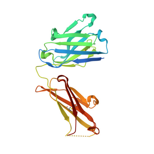
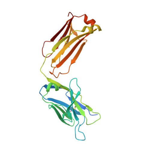
A straightforward approach to antibodies recognising cancer specific glycopeptidic neoepitopes

Wakui, H.Tanaka, Y.Ose, T.Matsumoto, I.Kato, K.Min, Y.Tachibana, T.Sato, M.Naruchi, K.Martin, F.G.Hinou, H.Nishimura, S.-I.

(2020) Chem Sci 11: 4999-5006

Macromolecule Content 

  • Total Structure Weight: 53.66 kDa 
  • Atom Count: 3,619 
  • Modeled Residue Count: 440 
  • Deposited Residue Count: 491 
  • Unique protein chains: 3

Macromolecules

Find similar proteins by:|  3D Structure
Entity ID: 1
MoleculeChains  Sequence LengthOrganismDetailsImage
Fab Fragment-SN-101-Heavy chain236Mus musculusMutation(s): 0 
Find similar proteins by:|  3D Structure
Entity ID: 2
MoleculeChains  Sequence LengthOrganismDetailsImage
Fab Fragment-SN-101-Light chain239Mus musculusMutation(s): 0 
Find similar proteins by:  Sequence   |   3D Structure  
Entity ID: 3
MoleculeChains  Sequence LengthOrganismDetailsImage
Synthetic MUC1 glycopepide16Homo sapiensMutation(s): 0 
UniProt & NIH Common Fund Data Resources
Find proteins for P15941 (Homo sapiens)
Explore P15941 
Go to UniProtKB:  P15941
PHAROS:  P15941
GTEx:  ENSG00000185499 
Entity Groups
Sequence Clusters30% Identity50% Identity70% Identity90% Identity95% Identity100% Identity
UniProt GroupP15941
Glycosylation
Glycosylation Sites: 1Go to GlyGen: P15941-1
Sequence Annotations
Expand
Reference Sequence

Small Molecules

Ligands 1 Unique
IDChains Name / Formula / InChI Key2D Diagram3D Interactions
A2G
(Subject of Investigation/LOI)

Query on A2G


Download:Ideal Coordinates CCD File
D [auth C]2-acetamido-2-deoxy-alpha-D-galactopyranose
C8 H15 N O6
OVRNDRQMDRJTHS-CBQIKETKSA-N

Experimental Data & Validation

Experimental Data

  • Method: X-RAY DIFFRACTION
  • Resolution: 1.77 Å
  • R-Value Free:  0.229 (Depositor), 0.230 (DCC) 
  • R-Value Work:  0.177 (Depositor), 0.179 (DCC) 
  • R-Value Observed: 0.179 (Depositor) 
Space Group: C 1 2 1
Unit Cell:
Length ( Å )Angle ( ˚ )
a = 159.92α = 90
b = 39.98β = 100.56
c = 63.87γ = 90
Software Package:
Software NamePurpose
PHENIXrefinement
XDSdata reduction
XDSdata scaling
PHASERphasing

Structure Validation

View Full Validation Report


Ligand Structure Quality Assessment 


Entry History 

& Funding Information

Deposition Data


Funding OrganizationLocationGrant Number
Japan Society for the Promotion of ScienceJapan25220206
Japan Society for the Promotion of ScienceJapan19H00918

Revision History  (Full details and data files)

  • Version 1.0: 2020-07-29
    Type: Initial release
  • Version 1.1: 2023-11-22
    Changes: Data collection, Database references, Refinement description, Structure summary
  • Version 1.2: 2024-11-20
    Changes: Structure summary