6KTK | pdb_00006ktk

Crystal structure of scyllo-inositol dehydrogenase R178A mutant, complexed with NADH and L-glucono-1,5-lactone, from Paracoccus laeviglucosivorans


Experimental Data Snapshot

  • Method: X-RAY DIFFRACTION
  • Resolution: 1.65 Å
  • R-Value Free: 
    0.171 (Depositor), 0.180 (DCC) 
  • R-Value Work: 
    0.160 (Depositor), 0.170 (DCC) 
  • R-Value Observed: 
    0.160 (Depositor) 

Starting Model: experimental
View more details

wwPDB Validation 3D Report Full Report

Validation slider image for 6KTK

Ligand Structure Quality Assessment 


This is version 1.4 of the entry. See complete history

Literature

Single amino acid mutation altered substrate specificity for L-glucose and inositol inscyllo-inositol dehydrogenase isolated fromParacoccus laeviglucosivorans.

Suzuki, M.Koubara, K.Takenoya, M.Fukano, K.Ito, S.Sasaki, Y.Nakamura, A.Yajima, S.

(2020) Biosci Biotechnol Biochem 84: 734-742

  • DOI: https://doi.org/10.1080/09168451.2019.1702870
  • Primary Citation Related Structures: 
    6KTJ, 6KTK, 6KTL

  • PubMed Abstract: 

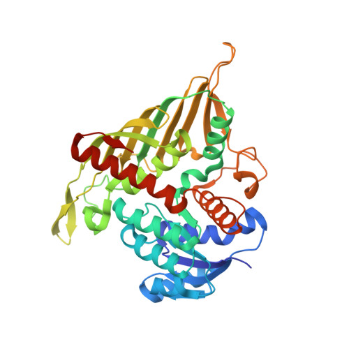
    scyllo- inositol dehydrogenase, isolated from Paracoccus laeviglucosivorans (Pl-sIDH), exhibits a broad substrate specificity: it oxidizes scyllo - and myo -inositols as well as L-glucose, converting L-glucose to L-glucono-1,5-lactone. Based on the crystal structures previously reported, Arg178 residue, located at the entry port of the catalytic site, seemed to be important for accepting substrates. Here, we report the role of Arg178 by using an alanine-substituted mutant for kinetic analysis as well as to determine the crystal structures. The wild-type Pl-sIDH exhibits the activity for scyllo -inositol most preferably followed by myo -inositol and L-glucose. On the contrary, the R178A mutant abolished the activities for both inositols, but remained active for L-glucose to the same extent as its wild-type. Based on the crystal structures of the mutant, the side chain of Asp191 flipped out of the substrate binding site. Therefore, Arg178 is important in positioning Asp191 correctly to exert its catalytic activities. Abbreviations: IDH: inositol dehydrogenase; LB: Luria-Bertani; k cat : catalyst rate constant; K m : Michaelis constant; NAD: nicotinamide dinucleotide; NADH: nicotinamide dinucleotide reduced form; PDB; Protein Data Bank; PDB entry: 6KTJ, 6KTK, 6KTL.


  • Organizational Affiliation
    • Department of Bioscience, Tokyo University of Agriculture, Tokyo, Japan.

Macromolecule Content 

  • Total Structure Weight: 169.03 kDa 
  • Atom Count: 12,419 
  • Modeled Residue Count: 1,469 
  • Deposited Residue Count: 1,520 
  • Unique protein chains: 1

Macromolecules

Find similar proteins by:|  3D Structure
Entity ID: 1
MoleculeChains  Sequence LengthOrganismDetailsImage
Scyllo-inositol dehydrogenase with L-glucose dehydrogenase activity
A, B, C, D
380Paracoccus laeviglucosivoransMutation(s): 1 
Gene Names: lgdA
UniProt
Find proteins for K7ZP76 (Paracoccus laeviglucosivorans)
Explore K7ZP76 
Go to UniProtKB:  K7ZP76
Entity Groups
Sequence Clusters30% Identity50% Identity70% Identity90% Identity95% Identity100% Identity
UniProt GroupK7ZP76
Sequence Annotations
Expand
Reference Sequence

Small Molecules

Ligands 3 Unique
IDChains Name / Formula / InChI Key2D Diagram3D Interactions
NAI

Query on NAI


Download:Ideal Coordinates CCD File
E [auth A],
H [auth B],
J [auth C],
L [auth D]
1,4-DIHYDRONICOTINAMIDE ADENINE DINUCLEOTIDE
C21 H29 N7 O14 P2
BOPGDPNILDQYTO-NNYOXOHSSA-N
PG4

Query on PG4


Download:Ideal Coordinates CCD File
F [auth A],
M [auth D]
TETRAETHYLENE GLYCOL
C8 H18 O5
UWHCKJMYHZGTIT-UHFFFAOYSA-N
8S0
(Subject of Investigation/LOI)

Query on 8S0


Download:Ideal Coordinates CCD File
G [auth A],
I [auth B],
K [auth C],
N [auth D]
L-glucono-1,5-lactone
C6 H10 O6
PHOQVHQSTUBQQK-KLVWXMOXSA-N

Experimental Data & Validation

Experimental Data

  • Method: X-RAY DIFFRACTION
  • Resolution: 1.65 Å
  • R-Value Free:  0.171 (Depositor), 0.180 (DCC) 
  • R-Value Work:  0.160 (Depositor), 0.170 (DCC) 
  • R-Value Observed: 0.160 (Depositor) 
Space Group: P 21 21 21
Unit Cell:
Length ( Å )Angle ( ˚ )
a = 90.61α = 90
b = 127.56β = 90
c = 137.57γ = 90
Software Package:
Software NamePurpose
REFMACrefinement
XDSdata reduction
XSCALEdata scaling
MOLREPphasing

Structure Validation

View Full Validation Report


Ligand Structure Quality Assessment 


Entry History 

& Funding Information

Deposition Data


Funding OrganizationLocationGrant Number
Japan Society for the Promotion of ScienceJapan16H04895

Revision History  (Full details and data files)

  • Version 1.0: 2019-12-25
    Type: Initial release
  • Version 1.1: 2020-01-01
    Changes: Database references
  • Version 1.2: 2020-03-04
    Changes: Database references
  • Version 1.3: 2020-07-29
    Type: Remediation
    Reason: Carbohydrate remediation
    Changes: Derived calculations, Structure summary
  • Version 1.4: 2023-11-22
    Changes: Data collection, Database references, Refinement description, Structure summary