


Heterochiral coupling in non-ribosomal peptide macrolactamization
Matsuda, K., Zhai, R., Mori, T., Kobayashi, M., Sano, A., Abe, I., Wakimoto, T.(2020) Nat Catal
Experimental Data Snapshot
Starting Model: experimental
View more details
(2020) Nat Catal
Entity ID: 1 | |||||
|---|---|---|---|---|---|
| Molecule | Chains | Sequence Length | Organism | Details | Image |
| Alpha/beta hydrolase | 471 | Streptomyces albidoflavus | Mutation(s): 0 | 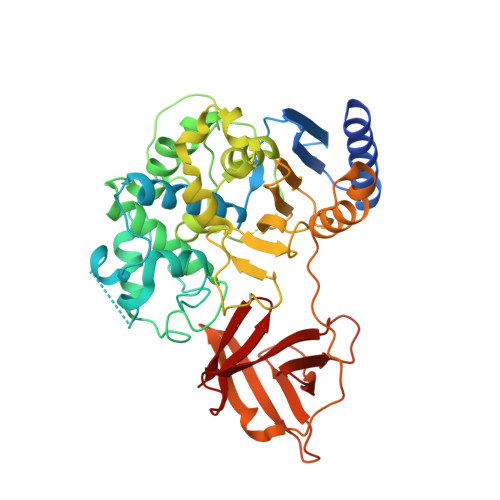 | |
UniProt | |||||
Find proteins for A0A679G4U8 (Streptomyces albidoflavus) Explore A0A679G4U8 Go to UniProtKB: A0A679G4U8 | |||||
Entity Groups | |||||
| Sequence Clusters | 30% Identity50% Identity70% Identity90% Identity95% Identity100% Identity | ||||
| UniProt Group | A0A679G4U8 | ||||
Sequence AnnotationsExpand | |||||
Reference Sequence | |||||
| Ligands 4 Unique | |||||
|---|---|---|---|---|---|
| ID | Chains | Name / Formula / InChI Key | 2D Diagram | 3D Interactions | |
| E7L (Subject of Investigation/LOI) Download:Ideal Coordinates CCD File | I [auth B] | S-(2-acetamidoethyl) (2R)-2-azanyl-4-methyl-pentanethioate C10 H20 N2 O2 S FGUKBCKKFXHYHQ-SECBINFHSA-N |  | ||
| DLE (Subject of Investigation/LOI) Download:Ideal Coordinates CCD File | C [auth A] | D-LEUCINE C6 H13 N O2 ROHFNLRQFUQHCH-RXMQYKEDSA-N |  | ||
| SO4 Download:Ideal Coordinates CCD File | D [auth A], E [auth A], F [auth A], J [auth B], K [auth B] | SULFATE ION O4 S QAOWNCQODCNURD-UHFFFAOYSA-L |  | ||
| YT3 Download:Ideal Coordinates CCD File | G [auth A], H [auth A], L [auth B] | YTTRIUM (III) ION Y GRTBAGCGDOYUBE-UHFFFAOYSA-N |  | ||
| Length ( Å ) | Angle ( ˚ ) |
|---|---|
| a = 48.37 | α = 90 |
| b = 152.06 | β = 113.08 |
| c = 64.54 | γ = 90 |
| Software Name | Purpose |
|---|---|
| XSCALE | data scaling |
| PHENIX | refinement |
| PDB_EXTRACT | data extraction |
| XDS | data reduction |
| PHASER | phasing |


