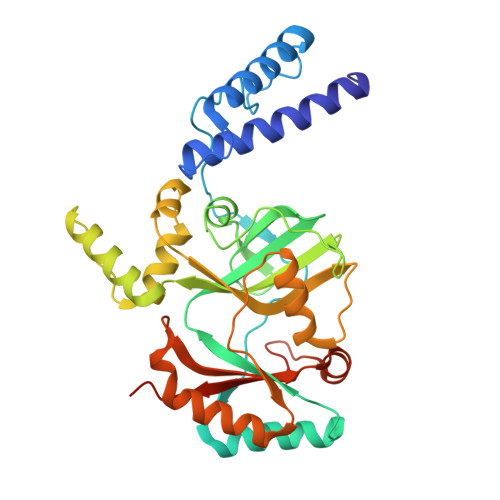
6KRH

Structural basis for domain rotation during adenylation of active site K123 and fragment library screening against NAD+ -dependent DNA ligase from Mycobacterium tuberculosis


Experimental Data Snapshot

  • Method: X-RAY DIFFRACTION
  • Resolution: 2.60 Å
  • R-Value Free: 0.264 
  • R-Value Work: 0.224 
  • R-Value Observed: 0.226 

Starting Model: experimental
View more details

wwPDB Validation   3D Report Full Report


Ligand Structure Quality Assessment 


This is version 1.2 of the entry. See complete history


Literature

Salt bridges at the subdomain interfaces of the adenylation domain and active-site residues of Mycobacterium tuberculosis NAD + -dependent DNA ligase A (MtbLigA) are important for the initial steps of nick-sealing activity.

Afsar, M.Shukla, A.Kumar, N.Ramachandran, R.

(2021) Acta Crystallogr D Struct Biol 77: 776-789

  • DOI: https://doi.org/10.1107/S2059798321003107
  • Primary Citation of Related Structures:  
    6KDU, 6KRH, 6KSC, 6KSD

  • PubMed Abstract: 

    NAD + -dependent DNA ligase (LigA) is the principal bacterial ligase and catalyses a multistep ligation reaction. The adenylation (AdD) domain at the N-terminus consists of subdomains 1a and 1b, where subdomain 1a is unique to LigA. Small-angle X-ray scattering and X-ray diffraction studies were used to probe changes in the relative spatial dispositions of the two subdomains during the adenylation reaction. Structural analyses of the inter-subdomain interactions of the AdD domain suggest that salt bridges formed by Glu22, Glu26 and Glu87 of subdomain 1a with Arg144, Arg315 and His240 of subdomain 1b play an important role in stabilizing the intermediate conformations of the two subdomains. E22A, E26A and E87A mutations reduce the in vitro activity by 89%, 64% and 39%, respectively, on a nicked DNA substrate, while they show no activity loss on a pre-adenylated DNA substrate, thus suggesting that the salt bridges are important in the initial steps of the ligation reaction. Furthermore, the E22A, E26A and E87A mutants exhibited extremely delayed growth in complementation assays involving the Escherichia coli GR501 strain, which harbours its own temperature-sensitive LigA. The H236A and H236Y mutants, which involve the residue that stacks against the adenine moiety of AMP, severely impact the activity and the ability to complement the growth-defective E. coli GR501 strain. Analysis of the K123A and K123R mutations in the active site rationalizes their total loss of activity and inability to rescue the growth-defective E. coli GR501 strain.


  • Organizational Affiliation

    Molecular and Structural Biology Division, CSIR - Central Drug Research Institute, Sector 10, Jankipuram Extension, Sitapur Road, Lucknow, Uttar Pradesh 226 031, India.


Macromolecules
Find similar proteins by:  (by identity cutoff)  |  3D Structure
Entity ID: 1
MoleculeChains Sequence LengthOrganismDetailsImage
DNA ligase A327Mycobacterium tuberculosis H37RvMutation(s): 1 
Gene Names: ligAligRv3014cMTV012.28c
EC: 6.5.1.2
UniProt
Find proteins for P9WNV1 (Mycobacterium tuberculosis (strain ATCC 25618 / H37Rv))
Explore P9WNV1 
Go to UniProtKB:  P9WNV1
Entity Groups  
Sequence Clusters30% Identity50% Identity70% Identity90% Identity95% Identity100% Identity
UniProt GroupP9WNV1
Sequence Annotations
Expand
  • Reference Sequence
Small Molecules
Ligands 3 Unique
IDChains Name / Formula / InChI Key2D Diagram3D Interactions
AMP (Subject of Investigation/LOI)
Query on AMP

Download Ideal Coordinates CCD File 
B [auth A]ADENOSINE MONOPHOSPHATE
C10 H14 N5 O7 P
UDMBCSSLTHHNCD-KQYNXXCUSA-N
NMN (Subject of Investigation/LOI)
Query on NMN

Download Ideal Coordinates CCD File 
C [auth A]BETA-NICOTINAMIDE RIBOSE MONOPHOSPHATE
C11 H16 N2 O8 P
DAYLJWODMCOQEW-TURQNECASA-O
SO4 (Subject of Investigation/LOI)
Query on SO4

Download Ideal Coordinates CCD File 
D [auth A],
E [auth A],
F [auth A],
G [auth A]
SULFATE ION
O4 S
QAOWNCQODCNURD-UHFFFAOYSA-L
Experimental Data & Validation

Experimental Data

  • Method: X-RAY DIFFRACTION
  • Resolution: 2.60 Å
  • R-Value Free: 0.264 
  • R-Value Work: 0.224 
  • R-Value Observed: 0.226 
  • Space Group: P 61 2 2
Unit Cell:
Length ( Å )Angle ( ˚ )
a = 95.549α = 90
b = 95.549β = 90
c = 200.589γ = 120
Software Package:
Software NamePurpose
PHENIXrefinement
Aimlessdata scaling
PDB_EXTRACTdata extraction
XDSdata reduction
PHASERphasing

Structure Validation

View Full Validation Report


Ligand Structure Quality Assessment 


Entry History 

Deposition Data

Revision History  (Full details and data files)

  • Version 1.0: 2020-08-26
    Type: Initial release
  • Version 1.1: 2021-06-09
    Changes: Database references
  • Version 1.2: 2023-11-22
    Changes: Data collection, Database references, Refinement description