


Uridine and triphosphate-bound UGPase from acinetobacter baumannii
Kang, L.W.To be published.
Experimental Data Snapshot
Starting Model: experimental
View more details
Entity ID: 1 | |||||
|---|---|---|---|---|---|
| Molecule | Chains | Sequence Length | Organism | Details | Image |
| UTP--glucose-1-phosphate uridylyltransferase | 290 | Acinetobacter baumannii | Mutation(s): 1 Gene Names: galU, B9X91_19205, CBI29_00108, CSB70_3798, DVA79_14980 EC: 2.7.7.9 | 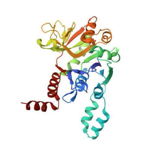 | |
UniProt | |||||
Entity Groups | |||||
| Sequence Clusters | 30% Identity50% Identity70% Identity90% Identity95% Identity100% Identity | ||||
| UniProt Group | X2KZJ9 | ||||
Sequence AnnotationsExpand | |||||
Reference Sequence | |||||
| Ligands 4 Unique | |||||
|---|---|---|---|---|---|
| ID | Chains | Name / Formula / InChI Key | 2D Diagram | 3D Interactions | |
| 3PO (Subject of Investigation/LOI) Download:Ideal Coordinates CCD File | D [auth A], F [auth B] | TRIPHOSPHATE H5 O10 P3 UNXRWKVEANCORM-UHFFFAOYSA-N |  | ||
| URI (Subject of Investigation/LOI) Download:Ideal Coordinates CCD File | C [auth A], E [auth B] | URIDINE C9 H12 N2 O6 DRTQHJPVMGBUCF-XVFCMESISA-N |  | ||
| SO4 Download:Ideal Coordinates CCD File | G [auth B], H [auth B] | SULFATE ION O4 S QAOWNCQODCNURD-UHFFFAOYSA-L |  | ||
| EDO Download:Ideal Coordinates CCD File | I [auth B] | 1,2-ETHANEDIOL C2 H6 O2 LYCAIKOWRPUZTN-UHFFFAOYSA-N |  | ||
| Length ( Å ) | Angle ( ˚ ) |
|---|---|
| a = 119.464 | α = 90 |
| b = 119.464 | β = 90 |
| c = 108.374 | γ = 90 |
| Software Name | Purpose |
|---|---|
| REFMAC | refinement |
| HKL-2000 | data scaling |
| HKL-2000 | data collection |
| HKL-2000 | data reduction |
| MOLREP | phasing |


