6KJU | pdb_00006kju

Huge conformation shift of Vibrio cholerae VqmA dimer in the absence of target DNA provides insight into DNA-binding mechanisms of LuxR-type receptors


Experimental Data Snapshot

  • Method: X-RAY DIFFRACTION
  • Resolution: 1.75 Å
  • R-Value Free: 
    0.196 (Depositor), 0.185 (DCC) 
  • R-Value Work: 
    0.175 (Depositor), 0.178 (DCC) 
  • R-Value Observed: 
    0.176 (Depositor) 

Starting Model: experimental
View more details

wwPDB Validation 3D Report Full Report

Validation slider image for 6KJU

Ligand Structure Quality Assessment 


This is version 1.2 of the entry. See complete history

Literature

Large conformation shifts of Vibrio cholerae VqmA dimer in the absence of target DNA provide insight into DNA-binding mechanisms of LuxR-type receptors.

Wu, H.Li, M.Peng, C.Yin, Y.Guo, H.Wang, W.Xu, Q.Zhou, H.Xu, C.Yu, F.He, J.

(2019) Biochem Biophys Res Commun 520: 399-405

  • DOI: https://doi.org/10.1016/j.bbrc.2019.10.063
  • Primary Citation Related Structures: 
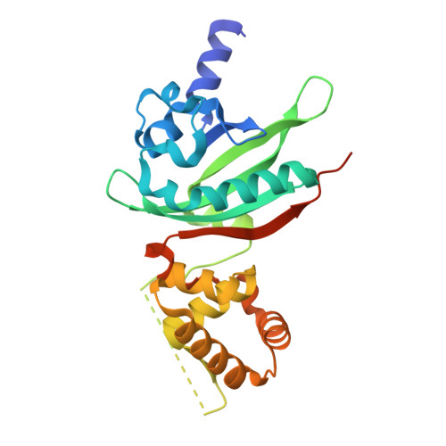
    6KJU

  • PubMed Abstract: 

    Quorum sensing regulates the biofilm formation and expression of virulence factors in Vibrio cholerae, an obligate human pathogen that continues to imperil human health. Cytoplasmic transcription factor VqmA is a LuxR-type receptor ubiquitous in the Vibrio genus and one vibriophage VP882 and plays an important role in V. cholerae pathogenicity. Here we presented the X-ray crystal structure of V. cholerae VqmA-DPO complex and compared it with the previously determined VqmA-DPO-DNA complex. To our knowledge, this is the first report on the crystal structures of the same LuxR-type receptor with two conformations of binding to DNA and not binding to DNA. Based on the results of structural analysis and biochemical assays, we revealed the secondary structure of the linker region between two function domains changed significantly, and DNA binding domains were covalently linked by a disulfide bond formed by the highly conserved Cys134. Besides, the distance between two DBD monomers became longer than that in DNA-binding conformation, and two α8 helixes underwent a large conformation shift. The results of the structure-function analyses presented here improve our understanding of the complex mechanisms in the conformational changes of LuxR-type receptors caused by DNA binding.


  • Organizational Affiliation
    • Shanghai Institute of Applied Physics, Chinese Academy of Sciences, Shanghai, 201800, China; University of Chinese Academy of Sciences, Beijing, 100049, China.

Macromolecule Content 

  • Total Structure Weight: 57.5 kDa 
  • Atom Count: 3,877 
  • Modeled Residue Count: 448 
  • Deposited Residue Count: 508 
  • Unique protein chains: 1

Macromolecules

Find similar proteins by:|  3D Structure
Entity ID: 1
MoleculeChains  Sequence LengthOrganismDetailsImage
Helix-turn-helix transcriptional regulator
A, B
254Vibrio choleraeMutation(s): 0 
Gene Names: C9J66_03310EC575_07960EN12_18880ERS013215_03763ET956_04375EYB64_07770
UniProt
Find proteins for Q9KKM5 (Vibrio cholerae serotype O1 (strain ATCC 39315 / El Tor Inaba N16961))
Explore Q9KKM5 
Go to UniProtKB:  Q9KKM5
Entity Groups
Sequence Clusters30% Identity50% Identity70% Identity90% Identity95% Identity100% Identity
UniProt GroupQ9KKM5
Sequence Annotations
Expand
Reference Sequence

Experimental Data & Validation

Experimental Data

  • Method: X-RAY DIFFRACTION
  • Resolution: 1.75 Å
  • R-Value Free:  0.196 (Depositor), 0.185 (DCC) 
  • R-Value Work:  0.175 (Depositor), 0.178 (DCC) 
  • R-Value Observed: 0.176 (Depositor) 
Space Group: P 21 21 21
Unit Cell:
Length ( Å )Angle ( ˚ )
a = 50.55α = 90
b = 84.51β = 90
c = 117.66γ = 90
Software Package:
Software NamePurpose
xia2data scaling
PHENIXrefinement
PDB_EXTRACTdata extraction
XDSdata reduction
PHENIXphasing

Structure Validation

View Full Validation Report


Ligand Structure Quality Assessment 


Entry History 

& Funding Information

Deposition Data


Funding OrganizationLocationGrant Number
National Science Foundation (China)China--

Revision History  (Full details and data files)

  • Version 1.0: 2019-11-13
    Type: Initial release
  • Version 1.1: 2023-11-22
    Changes: Data collection, Database references, Other, Refinement description
  • Version 1.2: 2024-10-16
    Changes: Structure summary