6KIP | pdb_00006kip

Crystal structure of PTPRD phosphatase domain in complex with Liprin-alpha3 tandem SAM domains


Experimental Data Snapshot

  • Method: X-RAY DIFFRACTION
  • Resolution: 1.91 Å
  • R-Value Free: 
    0.208 (Depositor), 0.209 (DCC) 
  • R-Value Work: 
    0.162 (Depositor), 0.169 (DCC) 
  • R-Value Observed: 
    0.165 (Depositor) 

Starting Models: experimental
View more details

wwPDB Validation 3D Report Full Report

Validation slider image for 6KIP

This is version 1.1 of the entry. See complete history

Literature

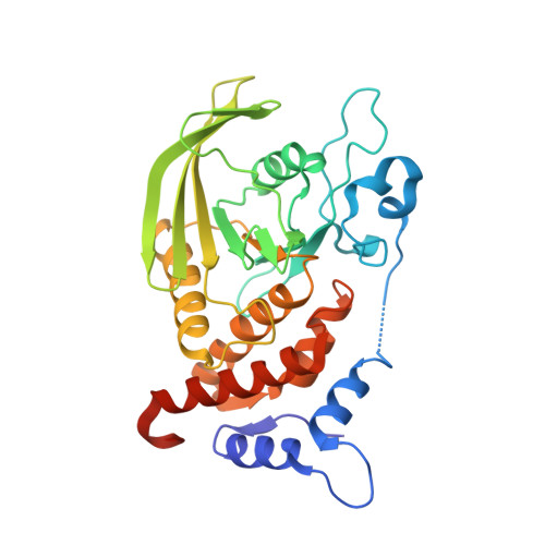
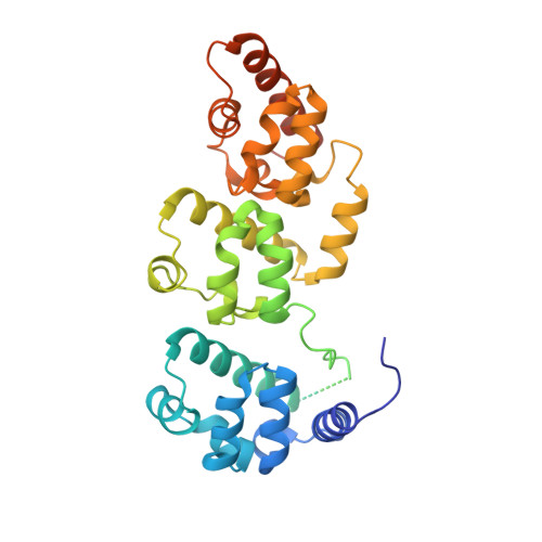
Structural insights into selective interaction between type IIa receptor protein tyrosine phosphatases and Liprin-alpha.

Wakita, M.Yamagata, A.Shiroshima, T.Izumi, H.Maeda, A.Sendo, M.Imai, A.Kubota, K.Goto-Ito, S.Sato, Y.Mori, H.Yoshida, T.Fukai, S.

(2020) Nat Commun 11: 649-649

  • DOI: https://doi.org/10.1038/s41467-020-14516-5
  • Primary Citation Related Structures: 
    6KIP

  • PubMed Abstract: 

    Synapse formation is induced by transsynaptic interaction of neuronal cell-adhesion molecules termed synaptic organizers. Type IIa receptor protein tyrosine phosphatases (IIa RPTPs) function as presynaptic organizers. The cytoplasmic domain of IIa RPTPs consists of two phosphatase domains, and the membrane-distal one (D2) is essential for synapse formation. Liprin-α, which is an active zone protein critical for synapse formation, interacts with D2 via its C-terminal domain composed of three tandem sterile alpha motifs (tSAM). Structural mechanisms of this critical interaction for synapse formation remain elusive. Here, we report the crystal structure of the complex between mouse PTPδ D2 and Liprin-α3 tSAM at 1.91 Å resolution. PTPδ D2 interacts with the N-terminal helix and the first and second SAMs (SAM1 and SAM2, respectively) of Liprin-α3. Structure-based mutational analyses in vitro and in cellulo demonstrate that the interactions with Liprin-α SAM1 and SAM2 are essential for the binding and synaptogenic activity.


  • Organizational Affiliation
    • Institute for Quantitative Biosciences, The University of Tokyo, Tokyo, 113-0032, Japan.

Macromolecule Content 

  • Total Structure Weight: 73.09 kDa 
  • Atom Count: 5,016 
  • Modeled Residue Count: 565 
  • Deposited Residue Count: 636 
  • Unique protein chains: 2

Macromolecules

Find similar proteins by:|  3D Structure
Entity ID: 1
MoleculeChains  Sequence LengthOrganismDetailsImage
Receptor-type tyrosine-protein phosphatase delta305Mus musculusMutation(s): 0 
Gene Names: Ptprd
EC: 3.1.3.48
UniProt & NIH Common Fund Data Resources
Find proteins for Q64487 (Mus musculus)
Explore Q64487 
Go to UniProtKB:  Q64487
IMPC:  MGI:97812
Entity Groups
Sequence Clusters30% Identity50% Identity70% Identity90% Identity95% Identity100% Identity
UniProt GroupQ64487
Sequence Annotations
Expand
Reference Sequence
Find similar proteins by:|  3D Structure
Entity ID: 2
MoleculeChains  Sequence LengthOrganismDetailsImage
Liprin-alpha-3331Mus musculusMutation(s): 0 
Gene Names: Ppfia3
UniProt & NIH Common Fund Data Resources
Find proteins for P60469 (Mus musculus)
Explore P60469 
Go to UniProtKB:  P60469
IMPC:  MGI:1924037
Entity Groups
Sequence Clusters30% Identity50% Identity70% Identity90% Identity95% Identity100% Identity
UniProt GroupP60469
Sequence Annotations
Expand
Reference Sequence

Experimental Data & Validation

Experimental Data

  • Method: X-RAY DIFFRACTION
  • Resolution: 1.91 Å
  • R-Value Free:  0.208 (Depositor), 0.209 (DCC) 
  • R-Value Work:  0.162 (Depositor), 0.169 (DCC) 
  • R-Value Observed: 0.165 (Depositor) 
Space Group: P 41 21 2
Unit Cell:
Length ( Å )Angle ( ˚ )
a = 98.012α = 90
b = 98.012β = 90
c = 140γ = 90
Software Package:
Software NamePurpose
HKL-2000data reduction
HKL-2000data scaling
MOLREPphasing
PHENIXrefinement
PDB_EXTRACTdata extraction

Structure Validation

View Full Validation Report


Entry History 

Deposition Data

Revision History  (Full details and data files)

  • Version 1.0: 2020-02-12
    Type: Initial release
  • Version 1.1: 2023-11-22
    Changes: Data collection, Database references, Refinement description