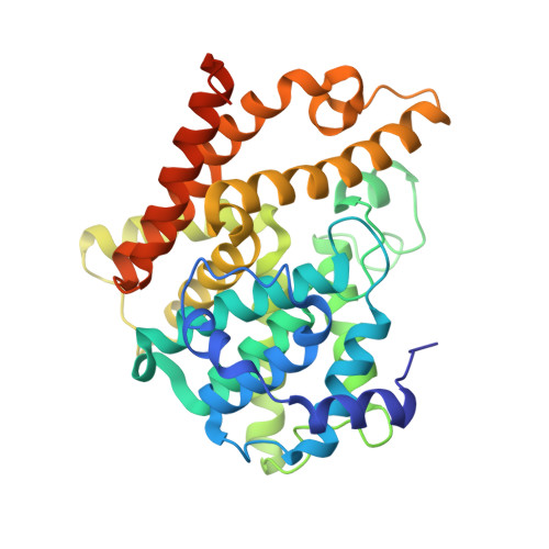
6KE0

Crystal structure of PDE10A in complex with a triazolopyrimidine inhibitor


Experimental Data Snapshot

  • Method: X-RAY DIFFRACTION
  • Resolution: 2.95 Å
  • R-Value Free: 0.302 
  • R-Value Work: 0.251 
  • R-Value Observed: 0.253 

Starting Model: experimental
View more details

wwPDB Validation   3D Report Full Report


Ligand Structure Quality Assessment 


This is version 1.1 of the entry. See complete history


Literature

Synthesis, SAR study, and biological evaluation of novel 2,3-dihydro-1H-imidazo[1,2-a]benzimidazole derivatives as phosphodiesterase 10A inhibitors.

Chino, A.Honda, S.Morita, M.Yonezawa, K.Hamaguchi, W.Amano, Y.Moriguchi, H.Yamazaki, M.Aota, M.Tomishima, M.Masuda, N.

(2019) Bioorg Med Chem 27: 3692-3706

  • DOI: https://doi.org/10.1016/j.bmc.2019.07.010
  • Primary Citation of Related Structures:  
    6KDX, 6KDZ, 6KE0

  • PubMed Abstract: 

    Phosphodiesterase 10A (PDE10A) inhibitors were designed and synthesized based on the dihydro-imidazobenzimidazole scaffold. Compound 5a showed moderate inhibitory activity and good permeability, but unfavorable high P-glycoprotein (P-gp) liability for brain penetration. We performed an optimization study to improve both the P-gp efflux ratio and PDE10A inhibitory activity. As a result, 6d was identified with improved P-gp liability and high PDE10A inhibitory activity. Compound 6d also showed satisfactory brain penetration, suppressed phencyclidine-induced hyperlocomotion and improved MK-801-induced working memory deficit.


  • Organizational Affiliation

    Functional Molecules, Modality Research Laboratories, Drug Discovery Research, Astellas Pharma Inc., 21, Miyukigaoka, Tsukuba, Ibaraki 305-8585, Japan. Electronic address: ayaka.chino@astellas.com.


Macromolecules
Find similar proteins by:  (by identity cutoff)  |  3D Structure
Entity ID: 1
MoleculeChains Sequence LengthOrganismDetailsImage
cAMP and cAMP-inhibited cGMP 3',5'-cyclic phosphodiesterase 10A
A, B
345Homo sapiensMutation(s): 0 
Gene Names: PDE10A
EC: 3.1.4.17 (PDB Primary Data), 3.1.4.35 (PDB Primary Data)
UniProt & NIH Common Fund Data Resources
Find proteins for Q9Y233 (Homo sapiens)
Explore Q9Y233 
Go to UniProtKB:  Q9Y233
PHAROS:  Q9Y233
GTEx:  ENSG00000112541 
Entity Groups  
Sequence Clusters30% Identity50% Identity70% Identity90% Identity95% Identity100% Identity
UniProt GroupQ9Y233
Sequence Annotations
Expand
  • Reference Sequence
Experimental Data & Validation

Experimental Data

  • Method: X-RAY DIFFRACTION
  • Resolution: 2.95 Å
  • R-Value Free: 0.302 
  • R-Value Work: 0.251 
  • R-Value Observed: 0.253 
  • Space Group: P 21 21 21
Unit Cell:
Length ( Å )Angle ( ˚ )
a = 50.398α = 90
b = 81.553β = 90
c = 160.763γ = 90
Software Package:
Software NamePurpose
HKL-2000data scaling
REFMACrefinement
PDB_EXTRACTdata extraction
HKL-2000data reduction
AMoREphasing

Structure Validation

View Full Validation Report


Ligand Structure Quality Assessment 


Entry History 

Deposition Data

Revision History  (Full details and data files)

  • Version 1.0: 2019-08-14
    Type: Initial release
  • Version 1.1: 2023-11-22
    Changes: Data collection, Database references, Derived calculations, Refinement description