6K97 | pdb_00006k97

Crystal structure of fusion DH domain


Experimental Data Snapshot

  • Method: X-RAY DIFFRACTION
  • Resolution: 2.50 Å
  • R-Value Free: 
    0.235 (Depositor), 0.215 (DCC) 
  • R-Value Work: 
    0.195 (Depositor) 
  • R-Value Observed: 
    0.197 (Depositor) 

Starting Model: experimental
View more details

wwPDB Validation 3D Report Full Report

Validation slider image for 6K97

This is version 1.2 of the entry. See complete history

Literature

Functional and Structural Analyses of the Split-Dehydratase Domain in the Biosynthesis of Macrolactam Polyketide Cremimycin.

Kawasaki, D.Miyanaga, A.Chisuga, T.Kudo, F.Eguchi, T.

(2019) Biochemistry 58: 4799-4803

  • DOI: https://doi.org/10.1021/acs.biochem.9b00897
  • Primary Citation Related Structures: 
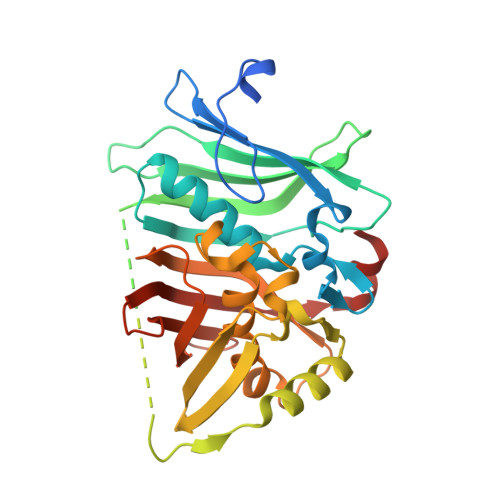
    6K97

  • PubMed Abstract: 

    In the biosynthesis of the macrolactam antibiotic cremimycin, the 3-aminononanoic acid starter unit is formed via a non-2-enoyl acyl carrier protein thioester intermediate, which is presumed to be constructed by cis -acyltransferase (AT) polyketide synthases (PKSs) CmiP2, CmiP3, and CmiP4. While canonical cis -AT PKS modules are comprised of a single polypeptide, the PKS module formed by CmiP2 and CmiP3 is split within the dehydratase (DH) domain. Here, we report the enzymatic function and the structural features of this split-DH domain. In vitro analysis showed that the split-DH domain catalyzes the dehydration reaction of ( R )-3-hydroxynonanoyl N -acetylcysteamine thioester (SNAC) to form ( E )-non-2-enoyl-SNAC, suggesting that the split-DH domain is catalytically active in cremimycin biosynthesis. In addition, structural analysis revealed that the CmiP2 and CmiP3 subunits of the split-DH domain form a tightly associated heterodimer through several hydrogen bonding and hydrophobic interactions, which are similar to those of canonical DH domains of other cis -AT PKSs. These results indicate that the split-DH domain has the same function and structure as common cis -AT PKS DH domains.


  • Organizational Affiliation
    • Department of Chemistry , Tokyo Institute of Technology , 2-12-1 O-okayama , Meguro-ku, Tokyo 152-8551 , Japan.

Macromolecule Content 

  • Total Structure Weight: 70.76 kDa 
  • Atom Count: 3,974 
  • Modeled Residue Count: 539 
  • Deposited Residue Count: 680 
  • Unique protein chains: 1

Macromolecules

Find similar proteins by:|  3D Structure
Entity ID: 1
MoleculeChains  Sequence LengthOrganismDetailsImage
Fusion DH
A, B
340Streptomyces sp. MJ635-86F5Mutation(s): 0 
Gene Names: cmiP3cmiP2
UniProt
Find proteins for X5IJ93 (Streptomyces sp. MJ635-86F5)
Explore X5IJ93 
Go to UniProtKB:  X5IJ93
Find proteins for X5IY86 (Streptomyces sp. MJ635-86F5)
Explore X5IY86 
Go to UniProtKB:  X5IY86
Entity Groups
Sequence Clusters30% Identity50% Identity70% Identity90% Identity95% Identity100% Identity
UniProt GroupsX5IY86X5IJ93
Sequence Annotations
Expand
Reference Sequence

Small Molecules

Ligands 1 Unique
IDChains Name / Formula / InChI Key2D Diagram3D Interactions
SO4

Query on SO4


Download:Ideal Coordinates CCD File
C [auth A]SULFATE ION
O4 S
QAOWNCQODCNURD-UHFFFAOYSA-L

Experimental Data & Validation

Experimental Data

  • Method: X-RAY DIFFRACTION
  • Resolution: 2.50 Å
  • R-Value Free:  0.235 (Depositor), 0.215 (DCC) 
  • R-Value Work:  0.195 (Depositor) 
  • R-Value Observed: 0.197 (Depositor) 
Space Group: C 1 2 1
Unit Cell:
Length ( Å )Angle ( ˚ )
a = 154.632α = 90
b = 66.564β = 90.97
c = 63.013γ = 90
Software Package:
Software NamePurpose
REFMACrefinement
XDSdata reduction
Aimlessdata scaling
MOLREPphasing

Structure Validation

View Full Validation Report


Entry History 

Deposition Data

Revision History  (Full details and data files)

  • Version 1.0: 2019-11-27
    Type: Initial release
  • Version 1.1: 2019-12-11
    Changes: Database references
  • Version 1.2: 2023-11-22
    Changes: Data collection, Database references, Refinement description