6K3A

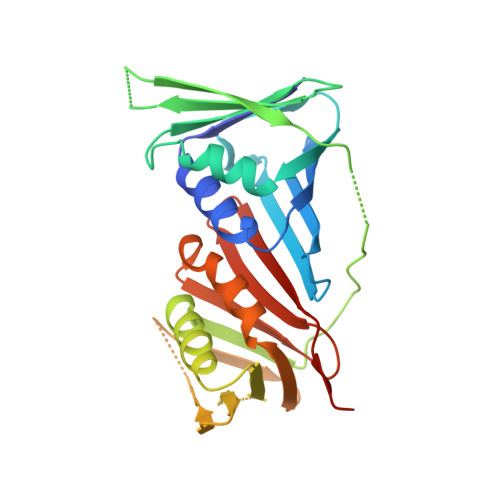
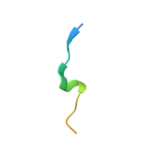
Crystal structure of human PCNA in complex with DNMT1 PIP box motif.

  • Classification: REPLICATION
  • Organism(s): Homo sapiens
  • Expression System: Escherichia coli
  • Mutation(s): No 

  • Deposited: 2019-05-17 Released: 2019-06-26 
  • Deposition Author(s): Jimenji, T., Kori, S., Arita, K.
  • Funding Organization(s): Ministry of Education, Culture, Sports, Science and Technology (Japan), Japan Science and Technology

Experimental Data Snapshot

  • Method: X-RAY DIFFRACTION
  • Resolution: 2.30 Å
  • R-Value Free: 0.264 
  • R-Value Work: 0.217 
  • R-Value Observed: 0.220 

Starting Model: experimental
View more details

wwPDB Validation   3D Report Full Report


This is version 1.3 of the entry. See complete history


Literature

Structure of PCNA in complex with DNMT1 PIP box reveals the basis for the molecular mechanism of the interaction.

Jimenji, T.Matsumura, R.Kori, S.Arita, K.

(2019) Biochem Biophys Res Commun 516: 578-583

  • DOI: https://doi.org/10.1016/j.bbrc.2019.06.060
  • Primary Citation of Related Structures:  
    6K3A

  • PubMed Abstract: 

    DNMT1 is a C5-DNA methyltransferase that plays a pivotal role in DNA methylation maintenance. During early and mid S-phase, DNMT1 accumulates at DNA replication sites by binding to proliferating cell nuclear antigen (PCNA), an essential factor for DNA replication, through a PIP box motif. However, the molecular mechanism by which the DNMT1 PIP box motif binds to PCNA remains unclear. Here, we report the crystal structure of PCNA bound to DNMT1 PIP box peptide. The structure reveals the detailed interaction between PCNA and DNMT1 PIP box; conserved glutamine and hydrophobic/aromatic residues in the PIP box are recognized by the Q- and hydrophobic pockets of PCNA, respectively. The structure also shows novel intramolecular interactions within the PIP box motif, which stabilize the helix conformation in the PIP box. Our data provide structural insight into the recruitment of DNMT1 to replication sites by PCNA.


  • Organizational Affiliation

    Structural Biology Laboratory, Graduate School of Medical Life Science, Yokohama City University, Yokohama, 230-0045, Japan.


Macromolecules
Find similar proteins by:  (by identity cutoff)  |  3D Structure
Entity ID: 1
MoleculeChains Sequence LengthOrganismDetailsImage
Proliferating cell nuclear antigen
A, C, E
264Homo sapiensMutation(s): 0 
Gene Names: PCNA
UniProt & NIH Common Fund Data Resources
Find proteins for P12004 (Homo sapiens)
Explore P12004 
Go to UniProtKB:  P12004
PHAROS:  P12004
GTEx:  ENSG00000132646 
Entity Groups  
Sequence Clusters30% Identity50% Identity70% Identity90% Identity95% Identity100% Identity
UniProt GroupP12004
Sequence Annotations
Expand
  • Reference Sequence

Find similar proteins by:  Sequence   |   3D Structure  

Entity ID: 2
MoleculeChains Sequence LengthOrganismDetailsImage
Peptide from DNA (cytosine-5)-methyltransferase 1
B, D, F
20Homo sapiensMutation(s): 0 
EC: 2.1.1.37
UniProt & NIH Common Fund Data Resources
Find proteins for P26358 (Homo sapiens)
Explore P26358 
Go to UniProtKB:  P26358
PHAROS:  P26358
GTEx:  ENSG00000130816 
Entity Groups  
Sequence Clusters30% Identity50% Identity70% Identity90% Identity95% Identity100% Identity
UniProt GroupP26358
Sequence Annotations
Expand
  • Reference Sequence
Experimental Data & Validation

Experimental Data

  • Method: X-RAY DIFFRACTION
  • Resolution: 2.30 Å
  • R-Value Free: 0.264 
  • R-Value Work: 0.217 
  • R-Value Observed: 0.220 
  • Space Group: C 1 2 1
Unit Cell:
Length ( Å )Angle ( ˚ )
a = 141.54α = 90
b = 81.598β = 90.02
c = 68.105γ = 90
Software Package:
Software NamePurpose
PHENIXrefinement
XDSdata reduction
Aimlessdata scaling
PHASERphasing

Structure Validation

View Full Validation Report


Entry History & Funding Information

Deposition Data


Funding OrganizationLocationGrant Number
Ministry of Education, Culture, Sports, Science and Technology (Japan)Japan18H02392
Japan Science and TechnologyJapan14530337

Revision History  (Full details and data files)

  • Version 1.0: 2019-06-26
    Type: Initial release
  • Version 1.1: 2019-07-10
    Changes: Data collection, Database references
  • Version 1.2: 2019-07-24
    Changes: Data collection, Database references
  • Version 1.3: 2023-11-22
    Changes: Data collection, Database references, Refinement description