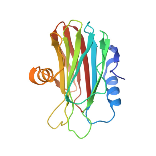
The Isolation of New Pore-Forming Toxins from the Sea AnemoneActinia fragaceaProvides Insights into the Mechanisms of Actinoporin Evolution.
Morante, K., Bellomio, A., Viguera, A.R., Gonzalez-Manas, J.M., Tsumoto, K., Caaveiro, J.M.M.(2019) Toxins (Basel) 11
- PubMed: 31295915
- DOI: https://doi.org/10.3390/toxins11070401
- Primary Citation of Related Structures:
6K2G - PubMed Abstract:
Random mutations and selective pressure drive protein adaptation to the changing demands of the environment. As a consequence, nature favors the evolution of protein diversity. A group of proteins subject to exceptional environmental stress and known for their widespread diversity are the pore-forming hemolytic proteins from sea anemones, known as actinoporins. In this study, we identified and isolated new isoforms of actinoporins from the sea anemone Actinia fragacea (fragaceatoxins). We characterized their hemolytic activity, examined their stability and structure, and performed a comparative analysis of their primary sequence. Sequence alignment reveals that most of the variability among actinoporins is associated with non-functional residues. The differences in the thermal behavior among fragaceatoxins suggest that these variability sites contribute to changes in protein stability. In addition, the protein-protein interaction region showed a very high degree of identity (92%) within fragaceatoxins, but only 25% among all actinoporins examined, suggesting some degree of specificity at the species level. Our findings support the mechanism of evolutionary adaptation in actinoporins and reflect common pathways conducive to protein variability.
- Department of Bioengineering, Graduate School of Engineering, The University of Tokyo, Bunkyo-ku, Tokyo 113-8656, Japan.
Organizational Affiliation:


