6K1H | pdb_00006k1h

Structure of membrane protein


Experimental Data Snapshot

  • Method: ELECTRON MICROSCOPY
  • Resolution: 3.52 Å
  • Aggregation State: PARTICLE 
  • Reconstruction Method: SINGLE PARTICLE 

wwPDB Validation 3D Report Full Report

Validation slider image for 6K1H

This is version 2.2 of the entry. See complete history

Literature

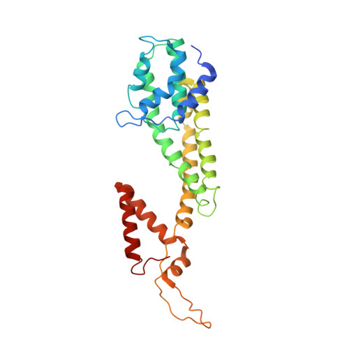
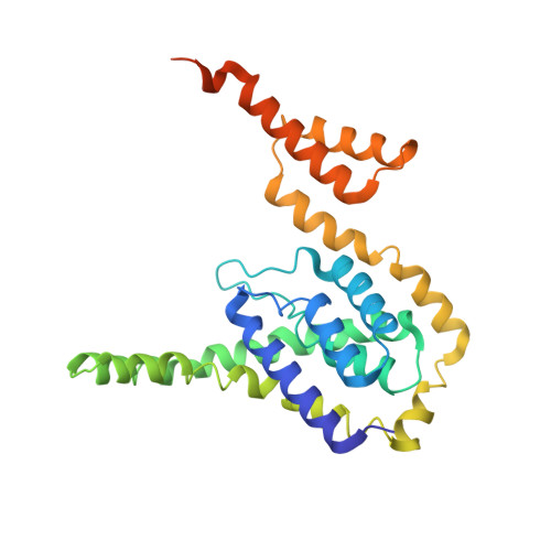
Structure of the mannose transporter of the bacterial phosphotransferase system.

Liu, X.Zeng, J.Huang, K.Wang, J.

(2019) Cell Res 29: 680-682

  • DOI: https://doi.org/10.1038/s41422-019-0194-z
  • Primary Citation Related Structures: 
    6K1H

  • Organizational Affiliation
    • State Key Laboratory of Membrane Biology, Beijing Advanced Innovation Center for Structural Biology, School of Life Sciences, Tsinghua University, 100084, Beijing, China.

Macromolecule Content 

  • Total Structure Weight: 179.75 kDa 
  • Atom Count: 11,769 
  • Modeled Residue Count: 1,560 
  • Deposited Residue Count: 1,671 
  • Unique protein chains: 2

Macromolecules

Find similar proteins by:|  3D Structure
Entity ID: 1
MoleculeChains  Sequence LengthOrganismDetailsImage
PTS mannose transporter subunit IIDA [auth Z],
C [auth F],
E [auth C]
283Escherichia coli str. K-12 substr. MG1655Mutation(s): 0 
Gene Names: D8B36_09735
Membrane Entity: Yes 
UniProt
Find proteins for P69805 (Escherichia coli (strain K12))
Explore P69805 
Go to UniProtKB:  P69805
Entity Groups
Sequence Clusters30% Identity50% Identity70% Identity90% Identity95% Identity100% Identity
UniProt GroupP69805
Sequence Annotations
Expand
Reference Sequence
Find similar proteins by:|  3D Structure
Entity ID: 2
MoleculeChains  Sequence LengthOrganismDetailsImage
PTS system mannose-specific EIIC componentB [auth Y],
D [auth E],
F [auth B]
274Escherichia coli K-12Mutation(s): 0 
Gene Names: manYpelptsPb1818JW1807
Membrane Entity: Yes 
UniProt
Find proteins for P69801 (Escherichia coli (strain K12))
Explore P69801 
Go to UniProtKB:  P69801
Entity Groups
Sequence Clusters30% Identity50% Identity70% Identity90% Identity95% Identity100% Identity
UniProt GroupP69801
Sequence Annotations
Expand
Reference Sequence

Experimental Data & Validation

Experimental Data

  • Method: ELECTRON MICROSCOPY
  • Resolution: 3.52 Å
  • Aggregation State: PARTICLE 
  • Reconstruction Method: SINGLE PARTICLE 
EM Software:
TaskSoftware PackageVersion
MODEL REFINEMENTPHENIX

Structure Validation

View Full Validation Report


Entry History 

& Funding Information

Deposition Data


Funding OrganizationLocationGrant Number
Ministry of Science and Technology (MoST, China)ChinaNo. 2016YFA0501103
Ministry of Science and Technology (MoST, China)China2015CB910104
National Natural Science Foundation of China (NSFC)China31621092

Revision History  (Full details and data files)

  • Version 1.0: 2019-07-10
    Type: Initial release
  • Version 1.1: 2019-07-17
    Changes: Data collection, Database references
  • Version 1.2: 2019-08-14
    Changes: Data collection, Database references
  • Version 1.3: 2020-07-29
    Type: Remediation
    Reason: Carbohydrate remediation
    Changes: Data collection, Derived calculations, Structure summary
  • Version 2.0: 2020-09-16
    Type: Coordinate replacement
    Reason: Model orientation/position
    Changes: Advisory, Atomic model, Author supporting evidence, Data collection, Database references, Derived calculations, Polymer sequence, Refinement description, Source and taxonomy, Structure summary
  • Version 2.1: 2024-03-27
    Changes: Data collection, Database references
  • Version 2.2: 2025-07-02
    Changes: Data collection, Structure summary