Development of a novel class of peroxisome proliferator-activated receptor (PPAR) gamma ligands as an anticancer agent with a unique binding mode based on a non-thiazolidinedione scaffold.
Yamamoto, K., Tamura, T., Nakamura, R., Hosoe, S., Matsubara, M., Nagata, K., Kodaira, H., Uemori, T., Takahashi, Y., Suzuki, M., Saito, J.I., Ueno, K., Shuto, S.(2019) Bioorg Med Chem 27: 115122-115122
- PubMed: 31623970 Search on PubMed
- DOI: https://doi.org/10.1016/j.bmc.2019.115122
- Primary Citation Related Structures:
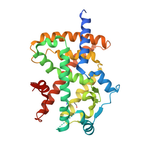
6K0T - PubMed Abstract:
We previously identified dibenzooxepine derivative 1 as a potent PPARγ ligand with a unique binding mode owing to its non-thiazolidinedione scaffold. However, while 1 showed remarkably potent MKN-45 gastric cancer cell aggregation activity, an indicator of cancer differentiation-inducing activity induced by PPARγ activation, we recognized that 1 was metabolically unstable. In the present study, we identified a metabolically soft spot, and successfully discovered 3-fluoro dibenzooxepine derivative 9 with better metabolic stability. Further optimization provided imidazo[1,2-a]pyridine derivative 17, which showed potent MKN-45 gastric cancer cell aggregation activity and excellent PK profiles compared with 9. Compound 17 exerted a growth inhibitory effect on AsPC-1/AG1 pancreatic tumor in mice. Furthermore, the decrease in the hematocrit (an indicator of localized edema, a serious adverse effect of PPARγ ligands) was tolerable even with oral administration at 200 mg/kg in healthy mice.
- Fuji Research Park, R&D Division, Kyowa Kirin, 1188, Shimotogari, Nagaizumi-cho, Sunto-gun, Shizuoka, Japan; Faculty of Pharmaceutical Sciences, Hokkaido University, Kita-12, Nishi-6, Kita-ku, Sapporo 060-0812, Japan. Electronic address: keisuke.yamamoto.fn@kyowakirin.com.
Organizational Affiliation: