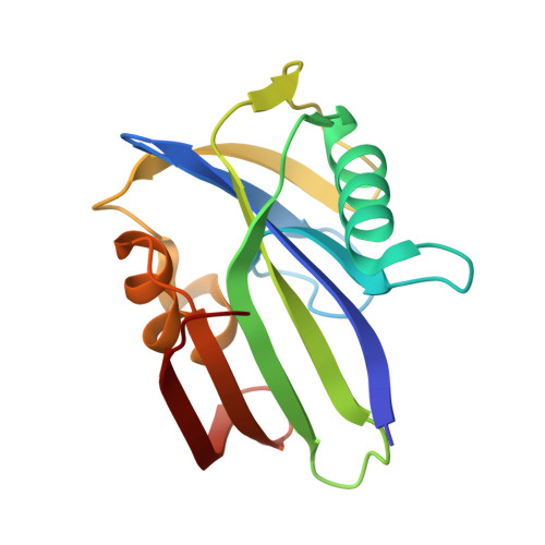
Inhibitor development of MTH1 via high-throughput screening with fragment based library and MTH1 substrate binding cavity.
Peng, C., Li, Y.H., Yu, C.W., Cheng, Z.H., Liu, J.R., Hsu, J.L., Hsin, L.W., Huang, C.T., Juan, H.F., Chern, J.W., Cheng, Y.S.(2021) Bioorg Chem 110: 104813-104813
- PubMed: 33774493
- DOI: https://doi.org/10.1016/j.bioorg.2021.104813
- Primary Citation of Related Structures:
6JVF, 6JVG, 6JVH, 6JVI, 6JVJ, 6JVK, 6JVL, 6JVM, 6JVN, 6JVO, 6JVP, 6JVQ, 6JVR, 6JVS, 6JVT - PubMed Abstract:
MutT Homolog 1 (MTH1) has been proven to hydrolyze oxidized nucleotide triphosphates during DNA repair. It can prevent the incorporation of wrong nucleotides during DNA replication and mitigate cell apoptosis. In a cancer cell, abundant reactive oxygen species can lead to substantial DNA damage and DNA mutations by base-pairing mismatch. MTH1 could eliminate oxidized dNTP and prevent cancer cells from entering cell death. Therefore, inhibition of MTH1 activity is considered to be an anti-cancer therapeutic target. In this study, high-throughput screening techniques were combined with a fragment-based library containing 2,313 compounds, which were used to screen for lead compounds with MTH1 inhibitor activity. Four compounds with MTH1 inhibitor ability were selected, and compound MI0639 was found to have the highest effective inhibition. To discover the selectivity and specificity of this action, several derivatives based on the MTH1 and MI0639 complex structure were synthesized. We compared 14 complex structures of MTH1 and the various compounds in combination with enzymatic inhibition and thermodynamic analysis. Nanomolar-range IC 50 inhibition abilities by enzyme kinetics and K d values by thermodynamic analysis were obtained for two compounds, named MI1020 and MI1024. Based on structural information and compound optimization, we aim to provide a strategy for the development of MTH1 inhibitors with high selectivity and specificity.
- Institute of Plant Biology, National Taiwan University, Taipei 10617, Taiwan.
Organizational Affiliation:


