


Decomposition of PET film by MHETase using Exo-PETase function
Sagong, H.-Y., Seo, H., Kim, T., Son, H., Joo, S., Lee, S., Kim, S., Woo, J.-S., Hwang, S., Kim, K.-J.(2020) ACS Catal 10: 4805-4812
Experimental Data Snapshot
wwPDB Validation 3D Report Full Report

(2020) ACS Catal 10: 4805-4812
Entity ID: 1 | |||||
|---|---|---|---|---|---|
| Molecule | Chains | Sequence Length | Organism | Details | Image |
| Mono(2-hydroxyethyl) terephthalate hydrolase | 613 | Pseudideonella sakaiensis | Mutation(s): 0 EC: 3.1.1.102 | 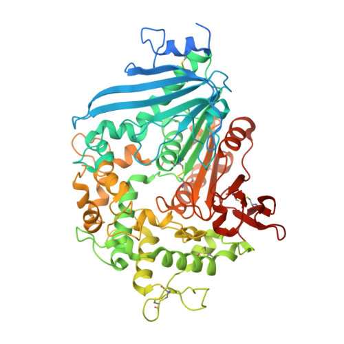 | |
UniProt | |||||
Find proteins for A0A0K8P8E7 (Piscinibacter sakaiensis) Explore A0A0K8P8E7 Go to UniProtKB: A0A0K8P8E7 | |||||
Entity Groups | |||||
| Sequence Clusters | 30% Identity50% Identity70% Identity90% Identity95% Identity100% Identity | ||||
| UniProt Group | A0A0K8P8E7 | ||||
Sequence AnnotationsExpand | |||||
Reference Sequence | |||||
| Ligands 5 Unique | |||||
|---|---|---|---|---|---|
| ID | Chains | Name / Formula / InChI Key | 2D Diagram | 3D Interactions | |
| GOL Download:Ideal Coordinates CCD File | E [auth A] K [auth B] L [auth B] M [auth B] N [auth B] | GLYCEROL C3 H8 O3 PEDCQBHIVMGVHV-UHFFFAOYSA-N |  | ||
| EDO Download:Ideal Coordinates CCD File | H [auth A], I [auth A] | 1,2-ETHANEDIOL C2 H6 O2 LYCAIKOWRPUZTN-UHFFFAOYSA-N |  | ||
| ACT Download:Ideal Coordinates CCD File | F [auth A], Q [auth B], V [auth C] | ACETATE ION C2 H3 O2 QTBSBXVTEAMEQO-UHFFFAOYSA-M |  | ||
| FMT Download:Ideal Coordinates CCD File | G [auth A] | FORMIC ACID C H2 O2 BDAGIHXWWSANSR-UHFFFAOYSA-N |  | ||
| CA (Subject of Investigation/LOI) Download:Ideal Coordinates CCD File | D [auth A], J [auth B], R [auth C] | CALCIUM ION Ca BHPQYMZQTOCNFJ-UHFFFAOYSA-N |  | ||
| Length ( Å ) | Angle ( ˚ ) |
|---|---|
| a = 214.931 | α = 90 |
| b = 173.163 | β = 119.86 |
| c = 107.225 | γ = 90 |
| Software Name | Purpose |
|---|---|
| HKL-2000 | data reduction |
| REFMAC | refinement |
| PDB_EXTRACT | data extraction |
| HKL-2000 | data scaling |
| PHASER | phasing |


