6JST | pdb_00006jst

Structure of Geobacillus kaustophilus lactonase, Y99P/D266N double mutant with bound 3-oxo-C8-HSL


Experimental Data Snapshot

  • Method: X-RAY DIFFRACTION
  • Resolution: 1.73 Å
  • R-Value Free: 
    0.264 (Depositor), 0.270 (DCC) 
  • R-Value Work: 
    0.219 (Depositor), 0.227 (DCC) 
  • R-Value Observed: 
    0.221 (Depositor) 

Starting Model: experimental
View more details

wwPDB Validation 3D Report Full Report

Validation slider image for 6JST

Ligand Structure Quality Assessment 


This is version 1.3 of the entry. See complete history

Literature

Directed Computational Evolution of Quorum-Quenching Lactonases from the Amidohydrolase Superfamily.

Go, M.K.Zhao, L.N.Xue, B.Supekar, S.Robinson, R.C.Fan, H.Yew, W.S.

(2020) Structure 28: 635

  • DOI: https://doi.org/10.1016/j.str.2020.03.011
  • Primary Citation Related Structures: 
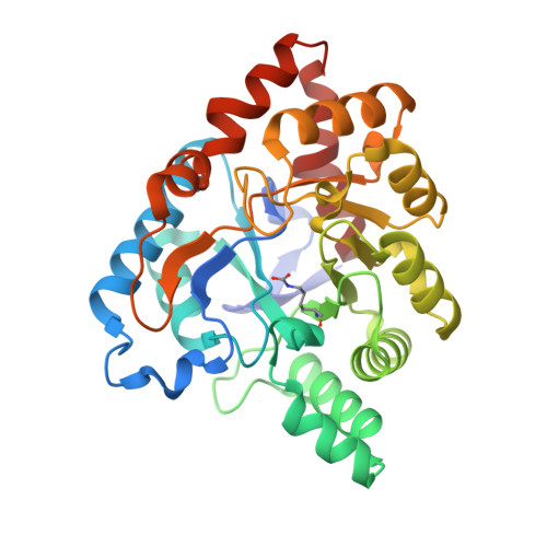
    6JSS, 6JST, 6JSU

  • PubMed Abstract: 

    In this work, we present a generalizable directed computational evolution protocol to effectively reduce the sequence space to be explored in rational enzyme design. The protocol involves in silico mutation modeling and substrate docking to rapidly identify mutagenesis hotspots that may enhance an enzyme's substrate binding and overall catalysis. By applying this protocol to a quorum-quenching Geobacillus kaustophilus lactonase, GKL, we generated 1,881 single mutants and docked high-energy intermediates of nine acyl homoserine lactones onto them. We found that Phe28 and Tyr99 were two hotspots that produced most of the predicted top 20 mutants. Of the 180 enzyme-substrate combinations (top 20 mutants × 9 substrates), 51 (28%) exhibited enhanced substrate binding and 22 (12%) had better overall activity when compared with wild-type GKL. X-ray crystallographic studies of Y99C and Y99P provided rationalized explanations for the enhancement in enzyme function and corroborated the utility of the protocol.


  • Organizational Affiliation
    • Department of Biochemistry, Yong Loo Lin School of Medicine, National University of Singapore, 8 Medical Drive, Singapore 117597, Singapore; NUS Synthetic Biology for Clinical and Technological Innovation, 14 Medical Drive, Singapore 117599, Singapore.

Macromolecule Content 

  • Total Structure Weight: 148.03 kDa 
  • Atom Count: 10,942 
  • Modeled Residue Count: 1,292 
  • Deposited Residue Count: 1,320 
  • Unique protein chains: 1

Macromolecules

Find similar proteins by:|  3D Structure
Entity ID: 1
MoleculeChains  Sequence LengthOrganismDetailsImage
Phosphotriesterase
A, B, C, D
330Geobacillus kaustophilus HTA426Mutation(s): 2 
Gene Names: GK1506
EC: 3.5
UniProt
Find proteins for Q5KZU5 (Geobacillus kaustophilus (strain HTA426))
Explore Q5KZU5 
Go to UniProtKB:  Q5KZU5
Entity Groups
Sequence Clusters30% Identity50% Identity70% Identity90% Identity95% Identity100% Identity
UniProt GroupQ5KZU5
Sequence Annotations
Expand
Reference Sequence

Small Molecules

Ligands 4 Unique
IDChains Name / Formula / InChI Key2D Diagram3D Interactions
LAE

Query on LAE


Download:Ideal Coordinates CCD File
H [auth A]3-OXO-OCTANOIC ACID (2-OXO-TETRAHYDRO-FURAN-3-YL)-AMIDE
C12 H19 N O4
FXCMGCFNLNFLSH-JTQLQIEISA-N
ZN

Query on ZN


Download:Ideal Coordinates CCD File
F [auth A],
J [auth B],
M [auth C],
P [auth D]
ZINC ION
Zn
PTFCDOFLOPIGGS-UHFFFAOYSA-N
FE

Query on FE


Download:Ideal Coordinates CCD File
E [auth A],
I [auth B],
L [auth C],
O [auth D]
FE (III) ION
Fe
VTLYFUHAOXGGBS-UHFFFAOYSA-N
OH

Query on OH


Download:Ideal Coordinates CCD File
G [auth A],
K [auth B],
N [auth C]
HYDROXIDE ION
H O
XLYOFNOQVPJJNP-UHFFFAOYSA-M
Modified Residues  1 Unique
IDChains TypeFormula2D DiagramParent
KCX
Query on KCX
A, B, C, D
L-PEPTIDE LINKINGC7 H14 N2 O4LYS

Experimental Data & Validation

Experimental Data

  • Method: X-RAY DIFFRACTION
  • Resolution: 1.73 Å
  • R-Value Free:  0.264 (Depositor), 0.270 (DCC) 
  • R-Value Work:  0.219 (Depositor), 0.227 (DCC) 
  • R-Value Observed: 0.221 (Depositor) 
Space Group: P 1
Unit Cell:
Length ( Å )Angle ( ˚ )
a = 50.991α = 90.99
b = 51.187β = 91.33
c = 134.473γ = 96.33
Software Package:
Software NamePurpose
XSCALEdata scaling
PHASERphasing
PHENIXrefinement
PDB_EXTRACTdata extraction
XDSdata reduction

Structure Validation

View Full Validation Report


Ligand Structure Quality Assessment 


Entry History 

& Funding Information

Deposition Data


Funding OrganizationLocationGrant Number
National Research Foundation (Singapore)Singapore--

Revision History  (Full details and data files)

  • Version 1.0: 2020-04-08
    Type: Initial release
  • Version 1.1: 2020-05-06
    Changes: Database references
  • Version 1.2: 2020-06-17
    Changes: Database references
  • Version 1.3: 2023-11-22
    Changes: Data collection, Database references, Derived calculations, Refinement description