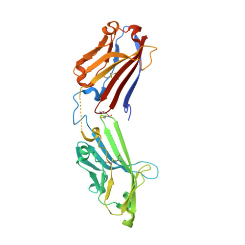
Arthritogenic alphaviruses, such as Chikungunya virus (CHIKV), cause severe and debilitating rheumatic diseases worldwide, resulting in severe morbidity and economic costs. Recently, MXRA8 was reported as an entry receptor. Here, we present the crystal structures of the mouse MXRA8, human MXRA8 in complex with the CHIKV E protein, and the cryo-electron microscopy structure of human MXRA8 and CHIKV virus-like particle. MXRA8 has two Ig-like domains with unique structural topologies. This receptor binds in the "canyon" between two protomers of the E spike on the surface of the virion. The atomic details at the interface between the two binding entities reveal that both the two domains and the hinge region of MXRA8 are involved in interaction with CHIKV E1-E2 residues from two protomers. Notably, the stalk region of MXRA8 is critical for CHIKV virus entry. This finding provides important information regarding the development of therapeutic countermeasures against those arthritogenic alphaviruses.
Organizational Affiliation:
Research Network of Immunity and Health (RNIH), Beijing Institutes of Life Science, Chinese Academy of Sciences, Beijing 100101, China.
CAS Key Laboratory of Pathogenic Microbiology and Immunology, Institute of Microbiology, Chinese Academy of Sciences, Beijing 100101, China; Savaid Medical School, University of Chinese Academy of Sciences, Beijing 100049, China.
CAS Key Laboratory of Pathogenic Microbiology and Immunology, Institute of Microbiology, Chinese Academy of Sciences, Beijing 100101, China.
CAS Key Laboratory of Pathogenic Microbiology and Immunology, Institute of Microbiology, Chinese Academy of Sciences, Beijing 100101, China; School of Life Sciences, University of Science and Technology of China, Hefei, Anhui 230026, China.
College of Veterinary Medicine, China Agricultural University, Beijing 100193, China.
Kunming Institute of Zoology, Chinese Academy of Sciences, Kunming 650223, China.
College of Animal Sciences and Technology, Guangxi University, Nanning 530004, China.
CAS Key Laboratory of Pathogenic Microbiology and Immunology, Institute of Microbiology, Chinese Academy of Sciences, Beijing 100101, China; National Institute of Science and Technology for Innovation on Diseases of Neglected Populations (INCT-IDPN), Center for Technological Development in Health (CDTS), Oswaldo Cruz Foundation (Fiocruz), Rio de Janeiro, Rio de Janeiro 21040-361, Brazil.
National Institute of Science and Technology for Innovation on Diseases of Neglected Populations (INCT-IDPN), Center for Technological Development in Health (CDTS), Oswaldo Cruz Foundation (Fiocruz), Rio de Janeiro, Rio de Janeiro 21040-361, Brazil.
CAS Key Laboratory of Pathogenic Microbiology and Immunology, Institute of Microbiology, Chinese Academy of Sciences, Beijing 100101, China; Savaid Medical School, University of Chinese Academy of Sciences, Beijing 100049, China; CAS Center for Influenza Research and Early-warning (CASCIRE), Chinese Academy of Sciences, Beijing 100101, China.
Tianjin Institute of Industrial Biotechnology, Chinese Academy of Sciences, Tianjin 300308, China; Institute of Genetics and Developmental Biology, Chinese Academy of Sciences, Beijing 100101, China. Electronic address: gaofeng@tib.cas.cn.
Research Network of Immunity and Health (RNIH), Beijing Institutes of Life Science, Chinese Academy of Sciences, Beijing 100101, China; CAS Key Laboratory of Pathogenic Microbiology and Immunology, Institute of Microbiology, Chinese Academy of Sciences, Beijing 100101, China; Savaid Medical School, University of Chinese Academy of Sciences, Beijing 100049, China; School of Life Sciences, University of Science and Technology of China, Hefei, Anhui 230026, China; CAS Center for Influenza Research and Early-warning (CASCIRE), Chinese Academy of Sciences, Beijing 100101, China; Tianjin Institute of Industrial Biotechnology, Chinese Academy of Sciences, Tianjin 300308, China; National Institute for Viral Disease Control and Prevention, Chinese Center for Disease Control and Prevention (China CDC), Beijing 102206, China. Electronic address: gaof@im.ac.cn.