


K3U bound crystal structure of class I type b peptide deformylase from Acinetobacter baumannii
Jung, K.H., Ho, T.H., Lee, I.H., Kang, L.W.To be published.
Experimental Data Snapshot
Starting Model: experimental
View more details
Entity ID: 1 | |||||
|---|---|---|---|---|---|
| Molecule | Chains | Sequence Length | Organism | Details | Image |
| Peptide deformylase | 159 | Acinetobacter baumannii | Mutation(s): 0 Gene Names: def, C3415_07350, IX87_00730 EC: 3.5.1.88 | 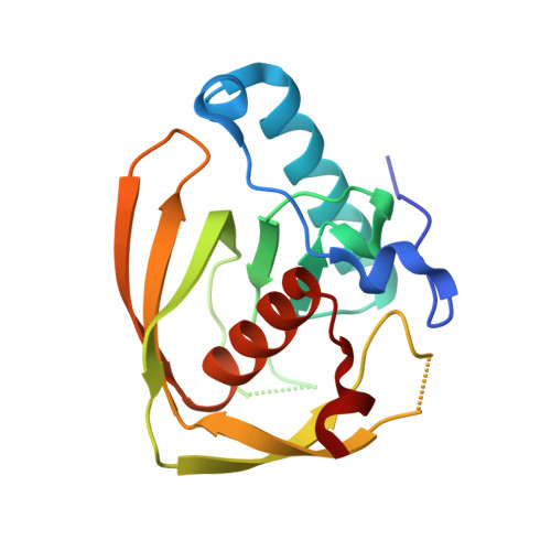 | |
| Ligands 2 Unique | |||||
|---|---|---|---|---|---|
| ID | Chains | Name / Formula / InChI Key | 2D Diagram | 3D Interactions | |
| K3U (Subject of Investigation/LOI) Download:Ideal Coordinates CCD File | D [auth A], F [auth B] | S-(2-oxo-2-phenylethyl) (2R)-2-benzyl-4,4,4-trifluorobutanethioate C19 H17 F3 O2 S CADIQQIULRUWPM-MRXNPFEDSA-N |  | ||
| ZN Download:Ideal Coordinates CCD File | C [auth A], E [auth B] | ZINC ION Zn PTFCDOFLOPIGGS-UHFFFAOYSA-N |  | ||
| Length ( Å ) | Angle ( ˚ ) |
|---|---|
| a = 39.513 | α = 90 |
| b = 71.259 | β = 90 |
| c = 110.974 | γ = 90 |
| Software Name | Purpose |
|---|---|
| HKL-2000 | data scaling |
| REFMAC | refinement |
| HKL-2000 | data collection |
| HKL-2000 | data reduction |
| MOLREP | phasing |


