


Crystal structure of GH10 family xylanase XynAF1 from Aspergillus fumigatus Z5
Li, G., Miao, Y., Zhang, R.To be published.
Experimental Data Snapshot
Entity ID: 1 | |||||
|---|---|---|---|---|---|
| Molecule | Chains | Sequence Length | Organism | Details | Image |
| Beta-xylanase | 319 | Aspergillus fumigatus Z5 | Mutation(s): 0 Gene Names: Y699_04481 EC: 3.2.1.8 | 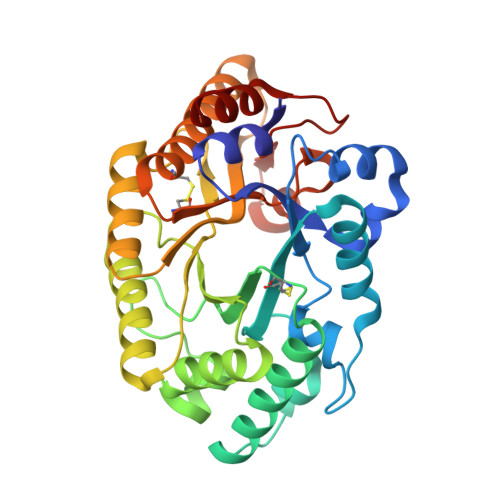 | |
| Ligands 2 Unique | |||||
|---|---|---|---|---|---|
| ID | Chains | Name / Formula / InChI Key | 2D Diagram | 3D Interactions | |
| NAG Download:Ideal Coordinates CCD File | C [auth A], E [auth B] | 2-acetamido-2-deoxy-beta-D-glucopyranose C8 H15 N O6 OVRNDRQMDRJTHS-FMDGEEDCSA-N |  | ||
| MES Download:Ideal Coordinates CCD File | D [auth A] | 2-(N-MORPHOLINO)-ETHANESULFONIC ACID C6 H13 N O4 S SXGZJKUKBWWHRA-UHFFFAOYSA-N |  | ||
| Length ( Å ) | Angle ( ˚ ) |
|---|---|
| a = 44.606 | α = 73.86 |
| b = 57.575 | β = 80.39 |
| c = 65.088 | γ = 68.35 |
| Software Name | Purpose |
|---|---|
| REFMAC | refinement |
| HKL-2000 | data reduction |
| HKL-2000 | data scaling |
| PDB_EXTRACT | data extraction |
| PHASER | phasing |


