6JDG

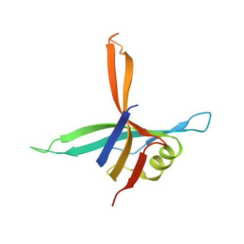
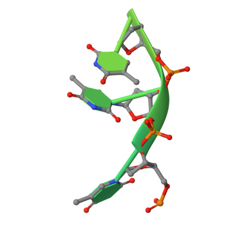
Complexed crystal structure of PaSSB with ssDNA dT20 at 2.39 angstrom resolution


Experimental Data Snapshot

  • Method: X-RAY DIFFRACTION
  • Resolution: 2.39 Å
  • R-Value Free: 0.264 
  • R-Value Work: 0.213 
  • R-Value Observed: 0.216 

wwPDB Validation   3D Report Full Report


This is version 1.1 of the entry. See complete history


Literature

Complexed crystal structure of SSB reveals a novel single-stranded DNA binding mode (SSB)3:1: Phe60 is not crucial for defining binding paths.

Huang, Y.H.Lin, E.S.Huang, C.Y.

(2019) Biochem Biophys Res Commun 520: 353-358

  • DOI: https://doi.org/10.1016/j.bbrc.2019.10.036
  • Primary Citation of Related Structures:  
    6JDG

  • PubMed Abstract: 

    Single-stranded DNA-binding protein (SSB) is essential to cells as it participates in DNA metabolic processes, such as DNA replication, repair, and recombination. Escherichia coli SSB (EcSSB) tetramer cooperatively binds and wraps ssDNA in two major binding modes. In this study, we report the complex crystal structure of Pseudomonas aeruginosa SSB (PaSSB) with ssDNA dT20 at 2.39 Å resolution (PDB entry 6JDG) that revealed a new binding mode, namely, (SSB) 3:1 . In the (SSB) 65 mode revealed by the EcSSB-dC35 complex structure, all four subunits fully participate in the binding to ssDNA. However, only three subunits in the PaSSB tetramer can participate in wrapping ssDNA in the (SSB) 3:1 mode. The bound ssDNA in the PaSSB-ssDNA complex adopts an Ω-shaped conformation rather than a χ-shaped conformation in the (SSB) 65 mode possibly due to the disability of Phe60. Phe60 is known to play a critical role in defining DNA-binding paths and promoting the wrapping of ssDNA around SSB tetramers. However, it is not important in the (SSB) 3:1 mode. The ssDNA binding path revealed by our structural evidence suggests that ssDNA occupies half of the binding sites of the two subunits and slightly comes into contact with the ssDNA binding sites of the third subunit. Accordingly, we propose and sketch a possible wrapping mechanism of SSB via this novel ssDNA-binding mode, (SSB) 3:1 .


  • Organizational Affiliation

    School of Biomedical Sciences, Chung Shan Medical University, No.110, Sec.1, Chien-Kuo N. Rd., Taichung City, Taiwan.


Macromolecules

Find similar proteins by:  (by identity cutoff)  |  3D Structure
Entity ID: 1
MoleculeChains Sequence LengthOrganismDetailsImage
Single-stranded DNA-binding protein
A, B, C, D
121Pseudomonas aeruginosa PAO1Mutation(s): 0 
Gene Names: ssbPA4232
UniProt
Find proteins for P40947 (Pseudomonas aeruginosa (strain ATCC 15692 / DSM 22644 / CIP 104116 / JCM 14847 / LMG 12228 / 1C / PRS 101 / PAO1))
Explore P40947 
Go to UniProtKB:  P40947
Entity Groups  
Sequence Clusters30% Identity50% Identity70% Identity90% Identity95% Identity100% Identity
UniProt GroupP40947
Sequence Annotations
Expand
  • Reference Sequence

Find similar nucleic acids by:  Sequence   |   3D Structure  

Entity ID: 2
MoleculeChains LengthOrganismImage
DNA (5'-D(P*TP*TP*TP*TP*TP*TP*TP*TP*TP*TP*TP*TP*TP*TP*TP*TP*TP*TP*TP*T)-3')
E, F, G
20synthetic construct
Sequence Annotations
Expand
  • Reference Sequence
Experimental Data & Validation

Experimental Data

  • Method: X-RAY DIFFRACTION
  • Resolution: 2.39 Å
  • R-Value Free: 0.264 
  • R-Value Work: 0.213 
  • R-Value Observed: 0.216 
  • Space Group: P 31
Unit Cell:
Length ( Å )Angle ( ˚ )
a = 60.042α = 90
b = 60.042β = 90
c = 129.642γ = 120
Software Package:
Software NamePurpose
PHENIXrefinement
HKL-2000data reduction
HKL-2000data scaling
MOLREPphasing

Structure Validation

View Full Validation Report


Entry History 

Deposition Data

Revision History  (Full details and data files)

  • Version 1.0: 2019-12-11
    Type: Initial release
  • Version 1.1: 2023-11-22
    Changes: Data collection, Database references, Refinement description