6JB1

Structure of pancreatic ATP-sensitive potassium channel bound with repaglinide and ATPgammaS at 3.3A resolution


Experimental Data Snapshot

  • Method: ELECTRON MICROSCOPY
  • Resolution: 3.30 Å
  • Aggregation State: PARTICLE 
  • Reconstruction Method: SINGLE PARTICLE 

wwPDB Validation   3D Report Full Report


This is version 2.0 of the entry. See complete history


Literature

The Structural Basis for the Binding of Repaglinide to the Pancreatic KATPChannel.

Ding, D.Wang, M.Wu, J.X.Kang, Y.Chen, L.

(2019) Cell Rep 27: 1848-1857.e4

  • DOI: https://doi.org/10.1016/j.celrep.2019.04.050
  • Primary Citation of Related Structures:  
    6JB1, 6JB3

  • PubMed Abstract: 

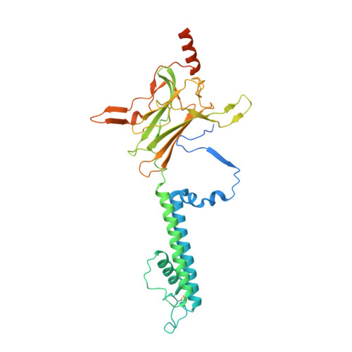
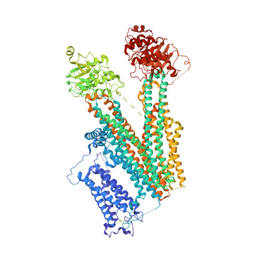
    Repaglinide (RPG) is a short-acting insulin secretagogue widely prescribed for the treatment of type 2 diabetes. It boosts insulin secretion by inhibiting the pancreatic ATP-sensitive potassium channel (K ATP ). However, the mechanisms by which RPG binds to the K ATP channel are poorly understood. Here, we describe two cryo-EM structures: the pancreatic K ATP channel in complex with inhibitory RPG and adenosine-5'-(γ-thio)-triphosphate (ATPγS) at 3.3 Å and a medium-resolution structure of a RPG-bound mini SUR1 protein in which the N terminus of the inward-rectifying potassium channel 6.1 (Kir6.1) is fused to the ABC transporter module of the sulfonylurea receptor 1 (SUR1). These structures reveal the binding site of RPG in the SUR1 subunit. Furthermore, the high-resolution structure reveals the complex architecture of the ATP binding site, which is formed by both Kir6.2 and SUR1 subunits, and the domain-domain interaction interfaces.


  • Organizational Affiliation

    State Key Laboratory of Membrane Biology, Institute of Molecular Medicine, Peking University, Beijing Key Laboratory of Cardiometabolic Molecular Medicine, Beijing 100871, China; Peking-Tsinghua Center for Life Sciences, Peking University, Beijing 100871, China; Academy for Advanced Interdisciplinary Studies, Peking University, Beijing 100871, China.


Macromolecules
Find similar proteins by:  (by identity cutoff)  |  3D Structure
Entity ID: 1
MoleculeChains Sequence LengthOrganismDetailsImage
ATP-sensitive inward rectifier potassium channel 11
A, C, E, G
390Mus musculusMutation(s): 0 
Gene Names: Kcnj11
Membrane Entity: Yes 
UniProt & NIH Common Fund Data Resources
Find proteins for Q61743 (Mus musculus)
Explore Q61743 
Go to UniProtKB:  Q61743
IMPC:  MGI:107501
Entity Groups  
Sequence Clusters30% Identity50% Identity70% Identity90% Identity95% Identity100% Identity
UniProt GroupQ61743
Sequence Annotations
Expand
  • Reference Sequence
Find similar proteins by:  (by identity cutoff)  |  3D Structure
Entity ID: 2
MoleculeChains Sequence LengthOrganismDetailsImage
ATP-binding cassette sub-family C member 8 isoform X2
B, D, F, H
1,582Mesocricetus auratusMutation(s): 0 
Gene Names: Abcc8
Membrane Entity: Yes 
UniProt
Find proteins for A0A1U7R319 (Mesocricetus auratus)
Explore A0A1U7R319 
Go to UniProtKB:  A0A1U7R319
Entity Groups  
Sequence Clusters30% Identity50% Identity70% Identity90% Identity95% Identity100% Identity
UniProt GroupA0A1U7R319
Sequence Annotations
Expand
  • Reference Sequence
Small Molecules
Ligands 5 Unique
IDChains Name / Formula / InChI Key2D Diagram3D Interactions
AJP
Query on AJP

Download Ideal Coordinates CCD File 
BA [auth D],
JB [auth H],
K [auth B],
SA [auth F]
Digitonin
C56 H92 O29
UVYVLBIGDKGWPX-KUAJCENISA-N
POV
Query on POV

Download Ideal Coordinates CCD File 
AA [auth C]
AB [auth F]
BB [auth F]
CB [auth F]
EA [auth D]
AA [auth C],
AB [auth F],
BB [auth F],
CB [auth F],
EA [auth D],
FA [auth D],
GA [auth D],
HA [auth D],
I [auth A],
IA [auth D],
IB [auth G],
JA [auth D],
KA [auth D],
LA [auth D],
MB [auth H],
N [auth B],
NB [auth H],
O [auth B],
OB [auth H],
P [auth B],
PB [auth H],
Q [auth B],
QB [auth H],
R [auth B],
RA [auth E],
RB [auth H],
S [auth B],
SB [auth H],
T [auth B],
TB [auth H],
U [auth B],
VA [auth F],
WA [auth F],
XA [auth F],
YA [auth F],
ZA [auth F]
(2S)-3-(hexadecanoyloxy)-2-[(9Z)-octadec-9-enoyloxy]propyl 2-(trimethylammonio)ethyl phosphate
C42 H82 N O8 P
WTJKGGKOPKCXLL-PFDVCBLKSA-N
PTY
Query on PTY

Download Ideal Coordinates CCD File 
DB [auth F]
EB [auth F]
FB [auth F]
GB [auth F]
MA [auth D]
DB [auth F],
EB [auth F],
FB [auth F],
GB [auth F],
MA [auth D],
NA [auth D],
OA [auth D],
PA [auth D],
UB [auth H],
V [auth B],
VB [auth H],
W [auth B],
WB [auth H],
X [auth B],
XB [auth H],
Y [auth B]
PHOSPHATIDYLETHANOLAMINE
C40 H80 N O8 P
NJGIRBISCGPRPF-KXQOOQHDSA-N
AGS
Query on AGS

Download Ideal Coordinates CCD File 
CA [auth D]
HB [auth G]
J [auth A]
KB [auth H]
L [auth B]
CA [auth D],
HB [auth G],
J [auth A],
KB [auth H],
L [auth B],
QA [auth E],
TA [auth F],
Z [auth C]
PHOSPHOTHIOPHOSPHORIC ACID-ADENYLATE ESTER
C10 H16 N5 O12 P3 S
NLTUCYMLOPLUHL-KQYNXXCUSA-N
BJX
Query on BJX

Download Ideal Coordinates CCD File 
DA [auth D],
LB [auth H],
M [auth B],
UA [auth F]
Repaglinide
C27 H36 N2 O4
FAEKWTJYAYMJKF-QHCPKHFHSA-N
Binding Affinity Annotations 
IDSourceBinding Affinity
BJX BindingDB:  6JB1 Kd: 50 (nM) from 1 assay(s)
IC50: 106 (nM) from 1 assay(s)
Experimental Data & Validation

Experimental Data

  • Method: ELECTRON MICROSCOPY
  • Resolution: 3.30 Å
  • Aggregation State: PARTICLE 
  • Reconstruction Method: SINGLE PARTICLE 
EM Software:
TaskSoftware PackageVersion
RECONSTRUCTIONRELION2.0

Structure Validation

View Full Validation Report


Entry History & Funding Information

Deposition Data


Funding OrganizationLocationGrant Number
Ministry of Science and Technology (China)China2016YFA0502004
National Natural Science Foundation of ChinaChina31622021
National Natural Science Foundation of ChinaChina31821091
National Natural Science Foundation of ChinaChina31870833

Revision History  (Full details and data files)

  • Version 1.0: 2019-05-22
    Type: Initial release
  • Version 1.1: 2019-11-06
    Changes: Data collection, Other
  • Version 2.0: 2021-09-29
    Changes: Advisory, Atomic model, Data collection, Database references