


Crystal structure of penta mutant of L-lactate dehydrogenase from Lactobacillus casei
Arai, K., Miyanaga, A., Uchikoba, H., Fushinobu, S., Taguchi, H.To be published.
Experimental Data Snapshot
Starting Model: experimental
View more details
Entity ID: 1 | |||||
|---|---|---|---|---|---|
| Molecule | Chains | Sequence Length | Organism | Details | Image |
| L-lactate dehydrogenase | 326 | Lacticaseibacillus casei DSM 20011 = JCM 1134 = ATCC 393 | Mutation(s): 0 Gene Names: ldh, LBCZ_2323 EC: 1.1.1.27 | 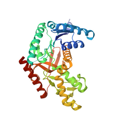 | |
UniProt | |||||
Entity Groups | |||||
| Sequence Clusters | 30% Identity50% Identity70% Identity90% Identity95% Identity100% Identity | ||||
| UniProt Group | P00343 | ||||
Sequence AnnotationsExpand | |||||
Reference Sequence | |||||
| Ligands 2 Unique | |||||
|---|---|---|---|---|---|
| ID | Chains | Name / Formula / InChI Key | 2D Diagram | 3D Interactions | |
| FBP Download:Ideal Coordinates CCD File | I [auth B], M [auth E], O [auth F] | 1,6-di-O-phosphono-beta-D-fructofuranose C6 H14 O12 P2 RNBGYGVWRKECFJ-ARQDHWQXSA-N |  | ||
| SO4 Download:Ideal Coordinates CCD File | G [auth A] H [auth B] J [auth C] K [auth D] L [auth E] | SULFATE ION O4 S QAOWNCQODCNURD-UHFFFAOYSA-L |  | ||
| Length ( Å ) | Angle ( ˚ ) |
|---|---|
| a = 164.25 | α = 90 |
| b = 83.582 | β = 91.53 |
| c = 180.094 | γ = 90 |
| Software Name | Purpose |
|---|---|
| REFMAC | refinement |
| HKL-2000 | data reduction |
| HKL-2000 | data scaling |
| MOLREP | phasing |


