6J4T

Crystal structure of arabidopsis ADAL complexed with IMP


Experimental Data Snapshot

  • Method: X-RAY DIFFRACTION
  • Resolution: 1.82 Å
  • R-Value Free: 0.229 
  • R-Value Work: 0.187 
  • R-Value Observed: 0.190 

wwPDB Validation   3D Report Full Report


Ligand Structure Quality Assessment 


This is version 1.2 of the entry. See complete history


Literature

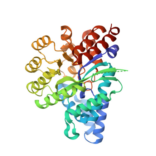
Structure ofArabidopsis thaliana N6-methyl-AMP deaminase ADAL with bound GMP and IMP and implications forN6-methyl-AMP recognition and processing.

Wu, B.Zhang, D.Nie, H.Shen, S.Li, Y.Li, S.

(2019) RNA Biol 16: 1504-1512

  • DOI: https://doi.org/10.1080/15476286.2019.1642712
  • Primary Citation of Related Structures:  
    6IV5, 6J23, 6J4T

  • PubMed Abstract: 

    Arabidopsis thaliana aminohydrolase ( At ADAL) has been shown to be involved in the metabolism of N 6 -methyl-AMP, a proposed intermediate during m 6 A-modified RNA metabolism, which can be subsequently incorporated into newly synthesized RNA by Pol II. It has been proposed that At ADAL will prevent N 6 -methyl-AMP reuse and catabolize it to inosine monophosphate (IMP). Here, we have solved the crystal structures of At ADAL in the apo form and in complex with GMP and IMP in the presence of Zn 2+ . We have identified the substrate-binding pocket of At ADAL and compared it with that for adenosine deaminase (ADA), adenine deaminase (ADE) and AMP deaminase (AMPD) from multiple species. The comparisons reveal that plant ADAL1 may have the potential ability to catalyze different alkyl-group substituted substrates.


  • Organizational Affiliation

    Department of Biology, Southern University of Science and Technology , Shenzhen , Guangdong , China.


Macromolecules
Find similar proteins by:  (by identity cutoff)  |  3D Structure
Entity ID: 1
MoleculeChains Sequence LengthOrganismDetailsImage
Adenosine/AMP deaminase family protein355Arabidopsis thalianaMutation(s): 1 
Gene Names: At4g04880T4B21.20T4B21_20
UniProt
Find proteins for Q8LPL7 (Arabidopsis thaliana)
Explore Q8LPL7 
Go to UniProtKB:  Q8LPL7
Entity Groups  
Sequence Clusters30% Identity50% Identity70% Identity90% Identity95% Identity100% Identity
UniProt GroupQ8LPL7
Sequence Annotations
Expand
  • Reference Sequence
Small Molecules
Experimental Data & Validation

Experimental Data

  • Method: X-RAY DIFFRACTION
  • Resolution: 1.82 Å
  • R-Value Free: 0.229 
  • R-Value Work: 0.187 
  • R-Value Observed: 0.190 
  • Space Group: P 21 21 21
Unit Cell:
Length ( Å )Angle ( ˚ )
a = 50.63α = 90
b = 81.921β = 90
c = 87.15γ = 90
Software Package:
Software NamePurpose
REFMACrefinement
iMOSFLMdata reduction
SCALAdata scaling
PHASERphasing

Structure Validation

View Full Validation Report


Ligand Structure Quality Assessment 


Entry History 

Deposition Data

Revision History  (Full details and data files)

  • Version 1.0: 2019-07-31
    Type: Initial release
  • Version 1.1: 2019-09-11
    Changes: Data collection, Database references
  • Version 1.2: 2023-11-22
    Changes: Data collection, Database references, Refinement description