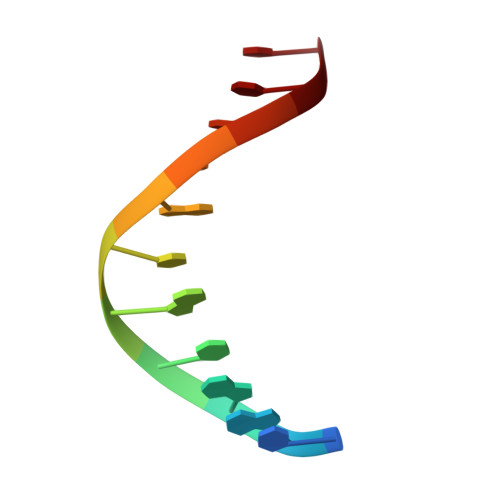
6J4R

Structural basis for the target DNA recognition and binding by the MYB domain of phosphate starvation response regulator 1


Experimental Data Snapshot

  • Method: X-RAY DIFFRACTION
  • Resolution: 2.80 Å
  • R-Value Free: 0.249 
  • R-Value Work: 0.215 
  • R-Value Observed: 0.217 

wwPDB Validation   3D Report Full Report


This is version 1.3 of the entry. See complete history


Literature

Structural basis for the Target DNA recognition and binding by the MYB domain of phosphate starvation response 1.

Jiang, M.Sun, L.Isupov, M.N.Littlechild, J.A.Wu, X.Wang, Q.Wang, Q.Yang, W.Wu, Y.

(2019) FEBS J 286: 2809-2821

  • DOI: https://doi.org/10.1111/febs.14846
  • Primary Citation of Related Structures:  
    6J4K, 6J4R, 6J5B

  • PubMed Abstract: 

    The phosphate starvation response 1 (PHR1) protein has a central role in mediating the response to phosphate starvation in plants. PHR1 is composed of a number of domains including a MYB domain involved with DNA binding and a coiled-coil domain proposed to be involved with dimer formation. PHR1 binds to the promoter of phosphate starvation-induced genes to control the levels of phosphate required for nutrition. Previous studies have shown that both the MYB domain and the coiled-coil domain of PHR1 are required for binding the target DNA. Here, we describe the crystal structure of the PHR1 MYB domain and two structures of its complex with the PHR1-binding DNA sequence (P1BS). Structural and isothermal titration calorimetry has been carried out showing that the MYB domain of PHR1 alone is sufficient for target DNA recognition and binding. Two copies of the PHR1 MYB domain bind to the same major groove of the P1BS DNA with few direct interactions between the individual MYB domains. In addition, the PHR1 MYB-P1BS DNA complex structures reveal amino acid residues involved in DNA recognition and binding. Mutagenesis of these residues results in lost or impaired ability of PHR1 MYB to bind to its target DNA. The results presented reveal the structural basis for DNA recognition by the PHR1 MYB domain and demonstrate that two PHR1 MYB domains attach to their P1BS DNA targeting sequence. DATABASE: Coordinates and structure factors have been deposited in the Protein Data Bank under accession codes 6J4K (PHR1 MYB), 6J4R (PHR1 MYB-R-P1BS), 6J5B (MYB-CC-R2-P1BS).


  • Organizational Affiliation

    State Key Laboratory of Structural Chemistry, Fujian Institute of Research on the Structure of Matter, Chinese Academy of Sciences, Fuzhou, China.


Macromolecules

Find similar proteins by:  (by identity cutoff)  |  3D Structure
Entity ID: 1
MoleculeChains Sequence LengthOrganismDetailsImage
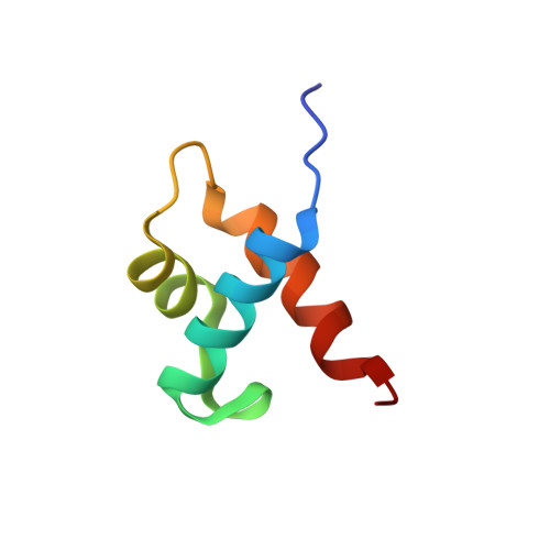
Protein PHOSPHATE STARVATION RESPONSE 1
A, B, C, D
58Arabidopsis thalianaMutation(s): 0 
Gene Names: PHR1At4g28610T5F17.60
UniProt
Find proteins for Q94CL7 (Arabidopsis thaliana)
Explore Q94CL7 
Go to UniProtKB:  Q94CL7
Entity Groups  
Sequence Clusters30% Identity50% Identity70% Identity90% Identity95% Identity100% Identity
UniProt GroupQ94CL7
Sequence Annotations
Expand
  • Reference Sequence

Find similar nucleic acids by:  Sequence   |   3D Structure  

Entity ID: 2
MoleculeChains LengthOrganismImage
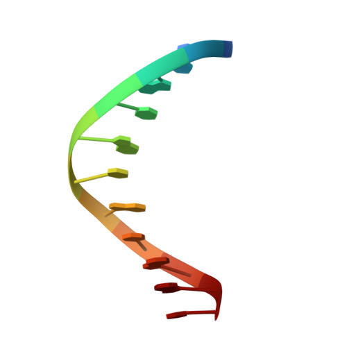
DNA (5'-D(*CP*CP*AP*TP*AP*TP*AP*TP*GP*AP*C)-3')E [auth F]11synthetic construct
Sequence Annotations
Expand
  • Reference Sequence

Find similar nucleic acids by:  Sequence   |   3D Structure  

Entity ID: 3
MoleculeChains LengthOrganismImage
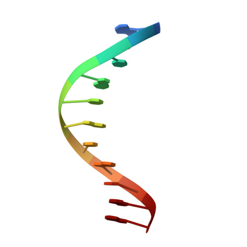
DNA (5'-D(*GP*TP*CP*AP*TP*AP*TP*AP*TP*GP*G)-3')F [auth E]11synthetic construct
Sequence Annotations
Expand
  • Reference Sequence

Find similar nucleic acids by:  Sequence   |   3D Structure  

Entity ID: 4
MoleculeChains LengthOrganismImage
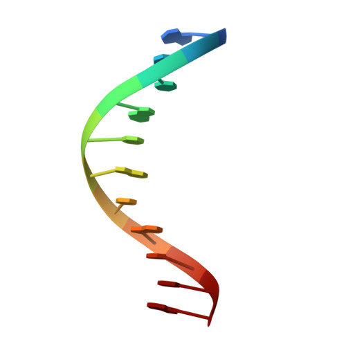
DNA (5'-D(*GP*GP*TP*AP*TP*AP*TP*AP*CP*TP*G)-3')G [auth X]11synthetic construct
Sequence Annotations
Expand
  • Reference Sequence

Find similar nucleic acids by:  Sequence   |   3D Structure  

Entity ID: 5
MoleculeChains LengthOrganismImage
DNA (5'-D(*CP*AP*GP*TP*AP*TP*AP*TP*AP*CP*C)-3')H [auth Y]11synthetic construct
Sequence Annotations
Expand
  • Reference Sequence
Experimental Data & Validation

Experimental Data

  • Method: X-RAY DIFFRACTION
  • Resolution: 2.80 Å
  • R-Value Free: 0.249 
  • R-Value Work: 0.215 
  • R-Value Observed: 0.217 
  • Space Group: P 42 21 2
Unit Cell:
Length ( Å )Angle ( ˚ )
a = 88.966α = 90
b = 88.966β = 90
c = 143.445γ = 90
Software Package:
Software NamePurpose
REFMACrefinement
xia2data scaling
PDB_EXTRACTdata extraction
xia2data reduction
PHENIXphasing

Structure Validation

View Full Validation Report


Entry History & Funding Information

Deposition Data


Funding OrganizationLocationGrant Number
National Science Foundation (China)China31470741

Revision History  (Full details and data files)

  • Version 1.0: 2019-04-24
    Type: Initial release
  • Version 1.1: 2019-07-31
    Changes: Data collection, Database references
  • Version 1.2: 2019-11-20
    Changes: Data collection
  • Version 1.3: 2024-03-27
    Changes: Data collection, Database references