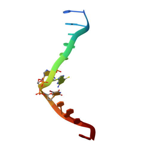
6IYQ | pdb_00006iyq

Crystal structure of a DNA duplex cross-linked by 6-thioguanine-6-thioguanine disulfides


Experimental Data Snapshot

  • Method: X-RAY DIFFRACTION
  • Resolution: 2.01 Å
  • R-Value Free: 
    0.282 (Depositor), 0.280 (DCC) 
  • R-Value Work: 
    0.247 (Depositor), 0.249 (DCC) 
  • R-Value Observed: 
    0.251 (Depositor) 

wwPDB Validation 3D Report Full Report

Validation slider image for 6IYQ

This is version 1.1 of the entry. See complete history. 

Literature

Crystal structure of a DNA duplex cross-linked by 6-thioguanine-6-thioguanine disulfides: reversible formation and cleavage catalyzed by Cu(II) ion and glutathione

Ono, A., Atsugi, T., Goto, M., Saneyoshi, H., Tomori, T., Seio, K., Dairaku, T., Kondo, J.

(2019) RSC Adv 

Macromolecule Content 

  • Total Structure Weight: 7.6 kDa 
  • Atom Count: 494 
  • Modeled Residue Count: 24 
  • Deposited Residue Count: 24 
  • Unique nucleic acid chains: 1

Macromolecules

Find similar nucleic acids by:  Sequence
Entity ID: 1
MoleculeChains LengthOrganismImage
DNA (5'-D(*CP*GP*CP*GP*AP*(S6G)P*(S6G)P*(BRU)P*CP*GP*CP*G)-3')
A, B
12synthetic construct
Sequence Annotations
Expand
Reference Sequence

Experimental Data & Validation

Experimental Data

  • Method: X-RAY DIFFRACTION
  • Resolution: 2.01 Å
  • R-Value Free:  0.282 (Depositor), 0.280 (DCC) 
  • R-Value Work:  0.247 (Depositor), 0.249 (DCC) 
  • R-Value Observed: 0.251 (Depositor) 
Space Group: I 2 2 2
Unit Cell:
Length ( Å )Angle ( ˚ )
a = 45.254α = 90
b = 46.522β = 90
c = 76.084γ = 90
Software Package:
Software NamePurpose
XSCALEdata scaling
SOLVEphasing
PHENIXrefinement
PDB_EXTRACTdata extraction
XDSdata reduction

Structure Validation

View Full Validation Report


Entry History 

Deposition Data

Revision History  (Full details and data files)

  • Version 1.0: 2019-10-30
    Type: Initial release
  • Version 1.1: 2024-03-27
    Changes: Data collection, Database references