6IYM | pdb_00006iym

Fumarylacetoacetate hydrolase (EaFAH) from psychrophilic Exiguobacterium antarcticum


Experimental Data Snapshot

  • Method: X-RAY DIFFRACTION
  • Resolution: 2.10 Å
  • R-Value Free: 
    0.269 (Depositor), 0.272 (DCC) 
  • R-Value Work: 
    0.202 (Depositor), 0.205 (DCC) 
  • R-Value Observed: 
    0.206 (Depositor) 

wwPDB Validation 3D Report Full Report

Validation slider image for 6IYM

This is version 1.1 of the entry. See complete history

Literature

Structural and functional analysis of a dimeric fumarylacetoacetate hydrolase (EaFAH) from psychrophilic Exiguobacterium antarcticum.

Yoo, W.Lee, C.W.Kim, B.Y.Huong Luu Le, L.T.Park, S.H.Kim, H.W.Shin, S.C.Kim, K.K.Lee, J.H.Kim, T.D.

(2019) Biochem Biophys Res Commun 509: 773-778

  • DOI: https://doi.org/10.1016/j.bbrc.2018.12.183
  • Primary Citation Related Structures: 
    6IYM

  • PubMed Abstract: 

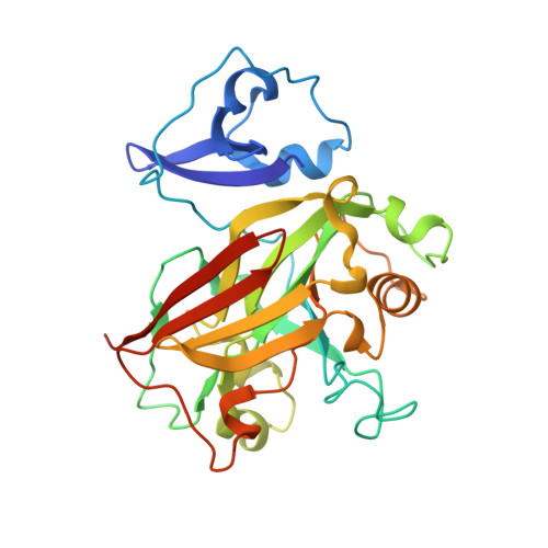
    Fumarylacetoacetate hydrolase (FAH) is essential for the degradation of aromatic amino acids as well as for the cleavage of carbon-carbon bonds in metabolites or small organic compounds. Here, the X-ray crystal structure of EaFAH, a dimeric fumarylacetoacetate hydrolase from Exiguobacterium antarcticum, was determined, and its functional properties were investigated using biochemical methods. EaFAH adopts a mixed β-sandwich roll fold with a highly flexible lid region (Val 73 -Leu 94 ), and an Mg 2+ ion is bound at the active site by coordinating to the three carboxylate oxygen atoms of Glu 124 , Glu 126 , and Asp 155 . The hydrolytic activity of EaFAH toward various substrates, including linalyl acetate was investigated using native polyacrylamide gel electrophoresis, activity staining, gel filtration, circular dichroism spectroscopy, fluorescence, and enzyme assays.


  • Organizational Affiliation
    • Department of Chemistry, College of Natural Science, Sookmyung Women's University, Seoul, 04310, South Korea; Department of Molecular Cell Biology, Samsung Biomedical Research Institute, Sungkyunkwan University School of Medicine, Suwon, 440-746, South Korea.

Macromolecule Content 

  • Total Structure Weight: 63.24 kDa 
  • Atom Count: 4,416 
  • Modeled Residue Count: 554 
  • Deposited Residue Count: 570 
  • Unique protein chains: 1

Macromolecules

Find similar proteins by:|  3D Structure
Entity ID: 1
MoleculeChains  Sequence LengthOrganismDetailsImage
5-oxopent-3-ene-1,2,5-tricarboxylate decarboxylase
A, B
285Exiguobacterium antarcticum B7Mutation(s): 0 
Gene Names: Eab7_1027

Experimental Data & Validation

Experimental Data

  • Method: X-RAY DIFFRACTION
  • Resolution: 2.10 Å
  • R-Value Free:  0.269 (Depositor), 0.272 (DCC) 
  • R-Value Work:  0.202 (Depositor), 0.205 (DCC) 
  • R-Value Observed: 0.206 (Depositor) 
Space Group: C 1 2 1
Unit Cell:
Length ( Å )Angle ( ˚ )
a = 120.437α = 90
b = 72.656β = 101.68
c = 63.374γ = 90
Software Package:
Software NamePurpose
REFMACrefinement
HKL-2000data reduction
HKL-2000data scaling
REFMACrefinement

Structure Validation

View Full Validation Report


Entry History 

Deposition Data

Revision History  (Full details and data files)

  • Version 1.0: 2019-04-03
    Type: Initial release
  • Version 1.1: 2024-03-27
    Changes: Data collection, Database references