6IWY | pdb_00006iwy

Crystal structure of the flagellar cap protein FliD from Helicobacter pylori


Experimental Data Snapshot

  • Method: X-RAY DIFFRACTION
  • Resolution: 2.60 Å
  • R-Value Free: 
    0.277 (Depositor), 0.271 (DCC) 
  • R-Value Work: 
    0.240 (Depositor), 0.239 (DCC) 
  • R-Value Observed: 
    0.242 (Depositor) 

wwPDB Validation 3D Report Full Report

Validation slider image for 6IWY

This is version 1.2 of the entry. See complete history

Literature

Structural analysis of the flagellar capping protein FliD from Helicobacter pylori.

Cho, S.Y.Song, W.S.Oh, H.B.Kim, H.U.Jung, H.S.Yoon, S.I.

(2019) Biochem Biophys Res Commun 514: 98-104

  • DOI: https://doi.org/10.1016/j.bbrc.2019.04.065
  • Primary Citation Related Structures: 
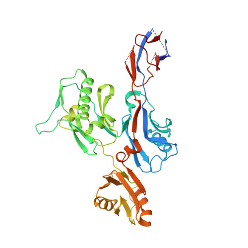
    6IWY

  • PubMed Abstract: 

    Helicobacter pylori is a pathogenic flagellated bacterium that infects the gastroduodenal mucosa and causes peptic ulcers in humans. FliD caps the distal end of the flagellar filament and is essential in filament growth. Moreover, FliD has been studied to diagnose and prevent H. pylori infection. Here, we report structure-based molecular studies of H. pylori FliD (hpFliD). A crystal structure of hpFliD at 2.6 Å resolution presents a four-domain (D2-D5) structure, where the D3 domain forms a central platform surrounded by the other three domains (D2, D4, and D5). hpFliD domains D2 and D3 structurally resemble those of FliD orthologs, whereas the D4 and D5 domains are exclusive to hpFliD. Moreover, our ELISA analysis using anti-H. pylori antibodies demonstrated that the hpFliD-specific D4 and D5 domains are highly antigenic compared to the D2 and D3 domains. Collectively, our structural and serological analyses underscore the structural role of hpFliD domains and provide a molecular basis for vaccine and diagnosis development.


  • Organizational Affiliation
    • Division of Biomedical Convergence, College of Biomedical Science, Kangwon National University, Chuncheon, 24341, Republic of Korea.

Macromolecule Content 

  • Total Structure Weight: 43.88 kDa 
  • Atom Count: 2,641 
  • Modeled Residue Count: 366 
  • Deposited Residue Count: 412 
  • Unique protein chains: 1

Macromolecules

Find similar proteins by:|  3D Structure
Entity ID: 1
MoleculeChains  Sequence LengthOrganismDetailsImage
Flagellar hook-associated protein 2412Helicobacter pylori 26695Mutation(s): 0 
Gene Names: fliDHP_0752
UniProt
Find proteins for P96786 (Helicobacter pylori (strain ATCC 700392 / 26695))
Explore P96786 
Go to UniProtKB:  P96786
Entity Groups
Sequence Clusters30% Identity50% Identity70% Identity90% Identity95% Identity100% Identity
UniProt GroupP96786
Sequence Annotations
Expand
Reference Sequence

Experimental Data & Validation

Experimental Data

  • Method: X-RAY DIFFRACTION
  • Resolution: 2.60 Å
  • R-Value Free:  0.277 (Depositor), 0.271 (DCC) 
  • R-Value Work:  0.240 (Depositor), 0.239 (DCC) 
  • R-Value Observed: 0.242 (Depositor) 
Space Group: P 2 2 21
Unit Cell:
Length ( Å )Angle ( ˚ )
a = 54.51α = 90
b = 75.891β = 90
c = 133.096γ = 90
Software Package:
Software NamePurpose
REFMACrefinement
HKL-2000data reduction
HKL-2000data scaling
PHENIXphasing

Structure Validation

View Full Validation Report


Entry History 

Deposition Data

Revision History  (Full details and data files)

  • Version 1.0: 2019-05-22
    Type: Initial release
  • Version 1.1: 2019-05-29
    Changes: Data collection, Database references
  • Version 1.2: 2024-03-27
    Changes: Data collection, Database references