6IWR | pdb_00006iwr

Crystal structure of GalNAc-T7 with UDP, GalNAc and Mn2+


Experimental Data Snapshot

  • Method: X-RAY DIFFRACTION
  • Resolution: 2.60 Å
  • R-Value Free: 
    0.258 (Depositor), 0.258 (DCC) 
  • R-Value Work: 
    0.220 (Depositor), 0.222 (DCC) 
  • R-Value Observed: 
    0.222 (Depositor) 

wwPDB Validation 3D Report Full Report

Validation slider image for 6IWR

Ligand Structure Quality Assessment 


This is version 1.3 of the entry. See complete history

Literature

Structural basis of carbohydrate transfer activity of UDP-GalNAc: Polypeptide N-acetylgalactosaminyltransferase 7.

Yu, C.Liang, L.Yin, Y.

(2019) Biochem Biophys Res Commun 510: 266-271

  • DOI: https://doi.org/10.1016/j.bbrc.2019.01.084
  • Primary Citation Related Structures: 
    6IWQ, 6IWR

  • PubMed Abstract: 

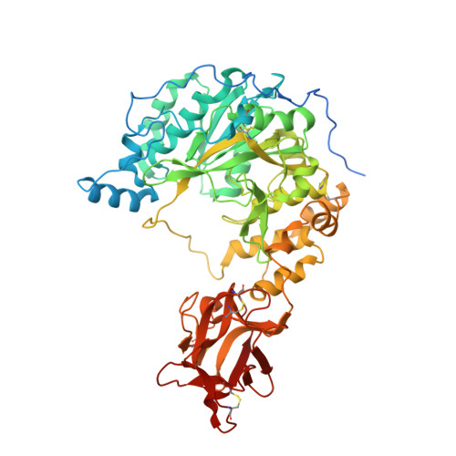
    The UDP-GalNAc:polypeptide N-acetylgalactosaminyltransferases (GalNAc-Ts) catalyze mucin-type O-glycosylation by transferring α-N-acetylgalactosamine (GalNAc) from UDP- GalNAc to Ser or Thr residues of target proteins. We resolved the crystal structures of GalNAc-T7, a GalNAc-T capable of glycosylating consecutive sites, and of its complex with the donor substrate UDP-GalNAc. The N-terminal catalytic domain and C-terminal lectin domain are connected by a flexible linker, forming a narrow cleft for the acceptor substrate. Only the α subdomain of the lectin domain binds to the glycosyl group, indicating that key residues determine substrate binding. Compared to the Apo structure, the loop covering the catalytic center of the complex show significant conformational changes, indicating the mechanism of the catalytic reaction.


  • Organizational Affiliation
    • Institute of Systems Biomedicine, School of Basic Medical Sciences, Peking University Health Science Center, Beijing, 100191, China; Department of Pathology, School of Basic Medical Sciences, Peking University Health Science Center, Beijing, 100191, China.

Macromolecule Content 

  • Total Structure Weight: 414.75 kDa 
  • Atom Count: 27,072 
  • Modeled Residue Count: 3,268 
  • Deposited Residue Count: 3,582 
  • Unique protein chains: 1

Macromolecules

Find similar proteins by:|  3D Structure
Entity ID: 1
MoleculeChains  Sequence LengthOrganismDetailsImage
N-acetylgalactosaminyltransferase 7
A, B, C, D, E
A, B, C, D, E, F
597Homo sapiensMutation(s): 0 
Gene Names: GALNT7
EC: 2.4.1 (PDB Primary Data), 2.4.1.41 (UniProt)
UniProt & NIH Common Fund Data Resources
Find proteins for Q86SF2 (Homo sapiens)
Explore Q86SF2 
Go to UniProtKB:  Q86SF2
PHAROS:  Q86SF2
GTEx:  ENSG00000109586 
Entity Groups
Sequence Clusters30% Identity50% Identity70% Identity90% Identity95% Identity100% Identity
UniProt GroupQ86SF2
Sequence Annotations
Expand
Reference Sequence

Small Molecules

Ligands 4 Unique
IDChains Name / Formula / InChI Key2D Diagram3D Interactions
UD2

Query on UD2


Download:Ideal Coordinates CCD File
M [auth D]URIDINE-DIPHOSPHATE-N-ACETYLGALACTOSAMINE
C17 H27 N3 O17 P2
LFTYTUAZOPRMMI-NESSUJCYSA-N
UDP

Query on UDP


Download:Ideal Coordinates CCD File
K [auth D]URIDINE-5'-DIPHOSPHATE
C9 H14 N2 O12 P2
XCCTYIAWTASOJW-XVFCMESISA-N
NAG

Query on NAG


Download:Ideal Coordinates CCD File
J [auth D]2-acetamido-2-deoxy-beta-D-glucopyranose
C8 H15 N O6
OVRNDRQMDRJTHS-FMDGEEDCSA-N
MN

Query on MN


Download:Ideal Coordinates CCD File
G [auth A]
H [auth B]
I [auth C]
L [auth D]
N [auth E]
G [auth A],
H [auth B],
I [auth C],
L [auth D],
N [auth E],
O [auth F]
MANGANESE (II) ION
Mn
WAEMQWOKJMHJLA-UHFFFAOYSA-N

Experimental Data & Validation

Experimental Data

  • Method: X-RAY DIFFRACTION
  • Resolution: 2.60 Å
  • R-Value Free:  0.258 (Depositor), 0.258 (DCC) 
  • R-Value Work:  0.220 (Depositor), 0.222 (DCC) 
  • R-Value Observed: 0.222 (Depositor) 
Space Group: P 21 21 21
Unit Cell:
Length ( Å )Angle ( ˚ )
a = 137.435α = 90
b = 158.264β = 90
c = 251.484γ = 90
Software Package:
Software NamePurpose
PHENIXrefinement
DENZOdata reduction
HKL-3000data scaling
PHASERphasing

Structure Validation

View Full Validation Report


Ligand Structure Quality Assessment 


Entry History 

Deposition Data

Revision History  (Full details and data files)

  • Version 1.0: 2019-02-06
    Type: Initial release
  • Version 1.1: 2019-02-20
    Changes: Data collection, Database references
  • Version 1.2: 2020-07-29
    Type: Remediation
    Reason: Carbohydrate remediation
    Changes: Data collection, Derived calculations, Structure summary
  • Version 1.3: 2024-11-20
    Changes: Data collection, Database references, Structure summary