6ITS | pdb_00006its

The citrate-bound trimer of chemoreceptor MCP2201 ligand binding domain


Experimental Data Snapshot

  • Method: X-RAY DIFFRACTION
  • Resolution: 2.50 Å
  • R-Value Free: 
    0.245 (Depositor), 0.247 (DCC) 
  • R-Value Work: 
    0.184 (Depositor), 0.188 (DCC) 
  • R-Value Observed: 
    0.190 (Depositor) 

Starting Model: experimental
View more details

wwPDB Validation 3D Report Full Report

Validation slider image for 6ITS

Ligand Structure Quality Assessment 


This is version 1.3 of the entry. See complete history

Literature

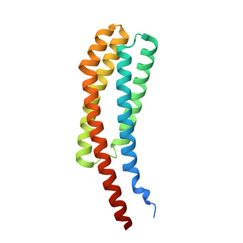
The ligand-binding domain of a chemoreceptor from Comamonas testosteroni has a previously unknown homotrimeric structure.

Hong, Y.Huang, Z.Guo, L.Ni, B.Jiang, C.Y.Li, X.J.Hou, Y.J.Yang, W.S.Wang, D.C.Zhulin, I.B.Liu, S.J.Li, D.F.

(2019) Mol Microbiol 112: 906-917

  • DOI: https://doi.org/10.1111/mmi.14326
  • Primary Citation Related Structures: 
    5XUA, 5XUB, 6ITS

  • PubMed Abstract: 

    Transmembrane chemoreceptors are widely present in Bacteria and Archaea. They play a critical role in sensing various signals outside and transmitting to the cell interior. Here, we report the structure of the periplasmic ligand-binding domain (LBD) of the transmembrane chemoreceptor MCP2201, which governs chemotaxis to citrate and other organic compounds in Comamonas testosteroni. The apo-form LBD crystal revealed a typical four-helix bundle homodimer, similar to previously well-studied chemoreceptors such as Tar and Tsr of Escherichia coli. However, the citrate-bound LBD revealed a four-helix bundle homotrimer that had not been observed in bacterial chemoreceptor LBDs. This homotrimer was further confirmed with size-exclusion chromatography, analytical ultracentrifugation and cross-linking experiments. The physiological importance of the homotrimer for chemotaxis was demonstrated with site-directed mutations of key amino acid residues in C. testosteroni mutants.


  • Organizational Affiliation
    • State Key Laboratory of Microbial Resources, Institute of Microbiology, Chinese Academy of Sciences, No. 1 Beichen West Road, Chaoyang District, Beijing, 100101, China.

Macromolecule Content 

  • Total Structure Weight: 36.66 kDa 
  • Atom Count: 2,317 
  • Modeled Residue Count: 293 
  • Deposited Residue Count: 330 
  • Unique protein chains: 1

Macromolecules

Find similar proteins by:|  3D Structure
Entity ID: 1
MoleculeChains  Sequence LengthOrganismDetailsImage
Methyl-accepting chemotaxis sensory transducer
A, B
165Comamonas testosteroni CNB-2Mutation(s): 0 
Gene Names: CtCNB1_2201

Experimental Data & Validation

Experimental Data

  • Method: X-RAY DIFFRACTION
  • Resolution: 2.50 Å
  • R-Value Free:  0.245 (Depositor), 0.247 (DCC) 
  • R-Value Work:  0.184 (Depositor), 0.188 (DCC) 
  • R-Value Observed: 0.190 (Depositor) 
Space Group: H 3
Unit Cell:
Length ( Å )Angle ( ˚ )
a = 48.86α = 90
b = 48.86β = 90
c = 376.152γ = 120
Software Package:
Software NamePurpose
PHENIXrefinement
iMOSFLMdata reduction
SCALAdata scaling
PHASERphasing

Structure Validation

View Full Validation Report


Ligand Structure Quality Assessment 


Entry History 

& Funding Information

Deposition Data


Funding OrganizationLocationGrant Number
National Natural Science Foundation of ChinaChina31870037

Revision History  (Full details and data files)

  • Version 1.0: 2018-12-26
    Type: Initial release
  • Version 1.1: 2019-07-10
    Changes: Data collection, Database references
  • Version 1.2: 2019-09-25
    Changes: Data collection, Database references
  • Version 1.3: 2023-11-22
    Changes: Data collection, Database references, Refinement description