6IS2 | pdb_00006is2

Crystal Structure of Staphylococcus aureus response regulator ArlR receiver domain in complex with Mg


Experimental Data Snapshot

  • Method: X-RAY DIFFRACTION
  • Resolution: 1.59 Å
  • R-Value Free: 
    0.188 (Depositor), 0.188 (DCC) 
  • R-Value Work: 
    0.155 (Depositor), 0.157 (DCC) 
  • R-Value Observed: 
    0.156 (Depositor) 

wwPDB Validation 3D Report Full Report

Validation slider image for 6IS2

This is version 1.3 of the entry. See complete history

Literature

Deciphering the activation and recognition mechanisms of Staphylococcus aureus response regulator ArlR.

Ouyang, Z.Zheng, F.Chew, J.Y.Pei, Y.Zhou, J.Wen, K.Han, M.Lemieux, M.J.Hwang, P.M.Wen, Y.

(2019) Nucleic Acids Res 47: 11418-11429

  • DOI: https://doi.org/10.1093/nar/gkz891
  • Primary Citation Related Structures: 
    6IS1, 6IS2, 6IS3, 6IS4

  • PubMed Abstract: 

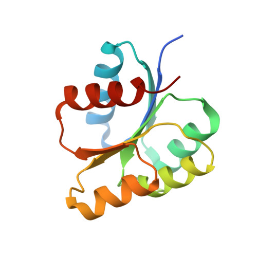
    Staphylococcus aureus ArlRS is a key two-component regulatory system necessary for adhesion, biofilm formation, and virulence. The response regulator ArlR consists of a C-terminal DNA-binding effector domain and an N-terminal receiver domain that is phosphorylated by ArlS, the cognate transmembrane sensor histidine kinase. We demonstrate that the receiver domain of ArlR adopts the canonical α5β5 response regulator assembly, which dimerizes upon activation, using beryllium trifluoride as an aspartate phosphorylation mimic. Activated ArlR recognizes a 20-bp imperfect inverted repeat sequence in the ica operon, which is involved in intercellular adhesion polysaccharide production. Crystal structures of the inactive and activated forms reveal that activation induces a significant conformational change in the β4-α4 and β5-α5-connecting loops, in which the α4 and α5 helices constitute the homodimerization interface. Crystal structures of the DNA-binding ArlR effector domain indicate that it is able to dimerize via a non-canonical β1-β2 hairpin domain swapping, raising the possibility of a new mechanism for signal transduction from the receiver domain to effector domain. Taken together, the current study provides structural insights into the activation of ArlR and its recognition, adding to the diversity of response regulation mechanisms that may inspire novel antimicrobial strategies specifically targeting Staphylococcus.


  • Organizational Affiliation
    • The Key Laboratory of Biomedical Information Engineering of Ministry of Education, School of Life Science and Technology, Department of Biochemistry and Molecular Biology, The Key Laboratory of Environment and Genes Related to Disease of Ministry of Education, Health Science Center, Xi'an Jiaotong University, Xi'an, China.

Macromolecule Content 

  • Total Structure Weight: 60.1 kDa 
  • Atom Count: 4,428 
  • Modeled Residue Count: 478 
  • Deposited Residue Count: 512 
  • Unique protein chains: 1

Macromolecules

Find similar proteins by:|  3D Structure
Entity ID: 1
MoleculeChains  Sequence LengthOrganismDetailsImage
Response regulator ArlR
A, B, C, D
128Staphylococcus aureus RF122Mutation(s): 0 
Gene Names: arlRSAB1271c
UniProt
Find proteins for Q2YY03 (Staphylococcus aureus (strain bovine RF122 / ET3-1))
Explore Q2YY03 
Go to UniProtKB:  Q2YY03
Entity Groups
Sequence Clusters30% Identity50% Identity70% Identity90% Identity95% Identity100% Identity
UniProt GroupQ2YY03
Sequence Annotations
Expand
Reference Sequence

Small Molecules

Ligands 4 Unique
IDChains Name / Formula / InChI Key2D Diagram3D Interactions
EPE

Query on EPE


Download:Ideal Coordinates CCD File
I [auth C]4-(2-HYDROXYETHYL)-1-PIPERAZINE ETHANESULFONIC ACID
C8 H18 N2 O4 S
JKMHFZQWWAIEOD-UHFFFAOYSA-N
SO4

Query on SO4


Download:Ideal Coordinates CCD File
F [auth A]SULFATE ION
O4 S
QAOWNCQODCNURD-UHFFFAOYSA-L
IMD

Query on IMD


Download:Ideal Coordinates CCD File
K [auth D]IMIDAZOLE
C3 H5 N2
RAXXELZNTBOGNW-UHFFFAOYSA-O
MG
(Subject of Investigation/LOI)

Query on MG


Download:Ideal Coordinates CCD File
E [auth A],
G [auth B],
H [auth C],
J [auth D]
MAGNESIUM ION
Mg
JLVVSXFLKOJNIY-UHFFFAOYSA-N

Experimental Data & Validation

Experimental Data

  • Method: X-RAY DIFFRACTION
  • Resolution: 1.59 Å
  • R-Value Free:  0.188 (Depositor), 0.188 (DCC) 
  • R-Value Work:  0.155 (Depositor), 0.157 (DCC) 
  • R-Value Observed: 0.156 (Depositor) 
Space Group: P 1
Unit Cell:
Length ( Å )Angle ( ˚ )
a = 43.589α = 99.72
b = 45.795β = 102.26
c = 61.847γ = 86.5
Software Package:
Software NamePurpose
PHENIXrefinement
XDSdata reduction
XDSdata scaling
MOLREPphasing

Structure Validation

View Full Validation Report


Entry History 

& Funding Information

Deposition Data


Funding OrganizationLocationGrant Number
National Natural Science Foundation of ChinaChinaNO.31870132, NO.81741088 and NO.31500051

Revision History  (Full details and data files)

  • Version 1.0: 2019-10-30
    Type: Initial release
  • Version 1.1: 2019-11-06
    Changes: Data collection, Derived calculations
  • Version 1.2: 2020-05-13
    Changes: Database references
  • Version 1.3: 2024-03-27
    Changes: Data collection, Database references