6INC | pdb_00006inc

Crystal structure of an acetolactate decarboxylase from Klebsiella pneumoniae


Experimental Data Snapshot

  • Method: X-RAY DIFFRACTION
  • Resolution: 1.60 Å
  • R-Value Free: 
    0.180 (Depositor), 0.181 (DCC) 
  • R-Value Work: 
    0.152 (Depositor), 0.153 (DCC) 
  • R-Value Observed: 
    0.154 (Depositor) 

Starting Model: experimental
View more details

wwPDB Validation 3D Report Full Report

Validation slider image for 6INC

This is version 1.1 of the entry. See complete history

Literature

Structural characterization of an acetolactate decarboxylase from Klebsiella pneumoniae

Wu, W.Zhao, Q.Che, S.Jia, H.Liang, H.Zhang, H.Liu, R.Zhang, Q.Bartlam, M.

(2019) Biochem Biophys Res Commun 509: 154-160

  • DOI: https://doi.org/10.1016/j.bbrc.2018.12.094
  • Primary Citation Related Structures: 
    6INB, 6INC

  • PubMed Abstract: 

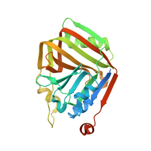
    Acetolactate decarboxylase (ALDC) is a well-characterized anabolic enzyme involved with 3-hydroxy butanone (acetoin), an important physiological metabolite excreted by microbes. Although the enzyme is widely present in microorganisms, few atomic structures and functions of ALDC have been reported to date. Here we report the crystal structure of ALDC from Klebsiella pneumoniae KP (KpALDC). KpALDC crystallizes in space group P3 1 21 with one monomer per asymmetric unit. Analytical ultracentrifugation data shows that KpALDC forms a stable dimer but can exist as a tetramer in solution. A Zn 2+ ion is coordinated by three strictly-conserved histidines (His198, His200 and His211) and a conserved glutamate (Glu69), but the C-terminal tail that forms part of the active site in ALDC enzymes is disordered. A complex structure with ethane-1,2-diol shows a unusual mode of binding, whereby the ligand does not coordinate the Zn 2+ ion. MicroScale Thermophoresis analysis shows that KpALDC binds Zn 2+ ions, but no binding of Mg 2+ , Ca 2+ and Mn 2+ ions was detected.


  • Organizational Affiliation
    • State Key Laboratory of Medicinal Chemical Biology, Nankai University, Tianjin, 300071, China; College of Life Sciences, Nankai University, Tianjin, 300071, China.

Macromolecule Content 

  • Total Structure Weight: 29.51 kDa 
  • Atom Count: 2,015 
  • Modeled Residue Count: 218 
  • Deposited Residue Count: 259 
  • Unique protein chains: 1

Macromolecules

Find similar proteins by:|  3D Structure
Entity ID: 1
MoleculeChains  Sequence LengthOrganismDetailsImage
Alpha-acetolactate decarboxylase259Klebsiella pneumoniaeMutation(s): 0 
Gene Names: budABN49_3166
EC: 4.1.1.5

Small Molecules

Ligands 4 Unique
IDChains Name / Formula / InChI Key2D Diagram3D Interactions
ZN

Query on ZN


Download:Ideal Coordinates CCD File
B [auth A]ZINC ION
Zn
PTFCDOFLOPIGGS-UHFFFAOYSA-N
EDO

Query on EDO


Download:Ideal Coordinates CCD File
E [auth A],
F [auth A]
1,2-ETHANEDIOL
C2 H6 O2
LYCAIKOWRPUZTN-UHFFFAOYSA-N
CL

Query on CL


Download:Ideal Coordinates CCD File
C [auth A],
D [auth A]
CHLORIDE ION
Cl
VEXZGXHMUGYJMC-UHFFFAOYSA-M
NA

Query on NA


Download:Ideal Coordinates CCD File
G [auth A]SODIUM ION
Na
FKNQFGJONOIPTF-UHFFFAOYSA-N

Experimental Data & Validation

Experimental Data

  • Method: X-RAY DIFFRACTION
  • Resolution: 1.60 Å
  • R-Value Free:  0.180 (Depositor), 0.181 (DCC) 
  • R-Value Work:  0.152 (Depositor), 0.153 (DCC) 
  • R-Value Observed: 0.154 (Depositor) 
Space Group: P 31 2 1
Unit Cell:
Length ( Å )Angle ( ˚ )
a = 55.152α = 90
b = 55.152β = 90
c = 119.377γ = 120
Software Package:
Software NamePurpose
PHENIXrefinement
HKL-3000data reduction
HKL-3000data scaling
PHASERphasing

Structure Validation

View Full Validation Report


Entry History 

& Funding Information

Deposition Data


Funding OrganizationLocationGrant Number
National Natural Science Foundation of ChinaChina31570128

Revision History  (Full details and data files)

  • Version 1.0: 2019-01-16
    Type: Initial release
  • Version 1.1: 2023-11-22
    Changes: Data collection, Database references, Refinement description